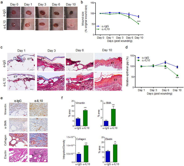
Figure 6. Blocking IL-10 signaling stimulates wound healing in diabetic wound.
db/db wounds were treated with either α-IgG or α-IL10 (10μg/wound) and assessed for wound healing by digital photography (a-b); by the histochemical assessment of re-epithelization and epidermal thickening, using H&E staining (c & d); and by histochemical assessments of the Vimentin, α-SMA, Masson’s Trichrome staining, and Elastin healing markers (e-f). Representative images are shown in (a, c, & e) and the corresponding data are plotted as mean ± SEM in (b, d, & f). Black scale bars=100μm, red scale bars=50μm. (N ≥ 5 mice/group, ≥9 random fields/wound/mouse, *p<0.05, ** p<0.01, ***p<0.001. Statistical analyses between groups were performed by One-way ANOVA, and pair-wise comparisons within groups were performed or by unpaired Student’s t-test).

