. 2022 Feb 17;63(3):229–240. doi: 10.3349/ymj.2022.63.3.229

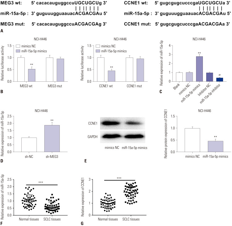
Fig. 6. Identification of a miR-15a-5p/CCNE1 axis as a downstream target of MEG3 in SCLC. (A) The predicted complementary binding sites between miR-15a-5p and MEG3/CCNE1. (B) The luciferase activity in NCI-H446 cells co-transfected with pGL3-MEG3 wt/pGL3-MEG3 mut and miR-15a-5p mimics/mimics NC or pGL3-CCNE1 wt/pGL3-CCNE1 mut and miR-15a-5p mimics/mimics NC was determined by dual luciferase reporter assay. **p<0.01 vs. the mimics NC group. (C) The expression of miR-15a-5p after transfection of miR-15a-5p mimics/mimics NC or miR-15a-5p inhibitor/inhibitor NC into NCI-H446 cells was detected by qRT-PCR. **p<0.01 vs. the mimics NC group, ##p<0.01 vs. the inhibitor NC group. (D) The expression of miR-15a-5p in NCI-H446 cells transfected with sh-MEG3/NC was detected by qRT-PCR. **p<0.01 vs. the sh-NC group. (E) The protein levels of CCNE1 in NCI-H446 cells transfected with miR-15a-5p mimics/mimics NC was determined by Western blot assay. **p<0.01 vs. the mimics NC group. (F) The expression of miR-15a-5p in SCLC tissues (n=48) and normal tissues (n=48) was detected by qRT-PCR. ***p<0.001 vs. the normal tissues. (G) The expression of CCNE1 in SCLC tissues (n=48) and normal tissues (n=48) was detected by qRT-PCR. ***p<0.001 vs. the normal tissues. SCLC, small-cell lung cancer.

Fig. 6