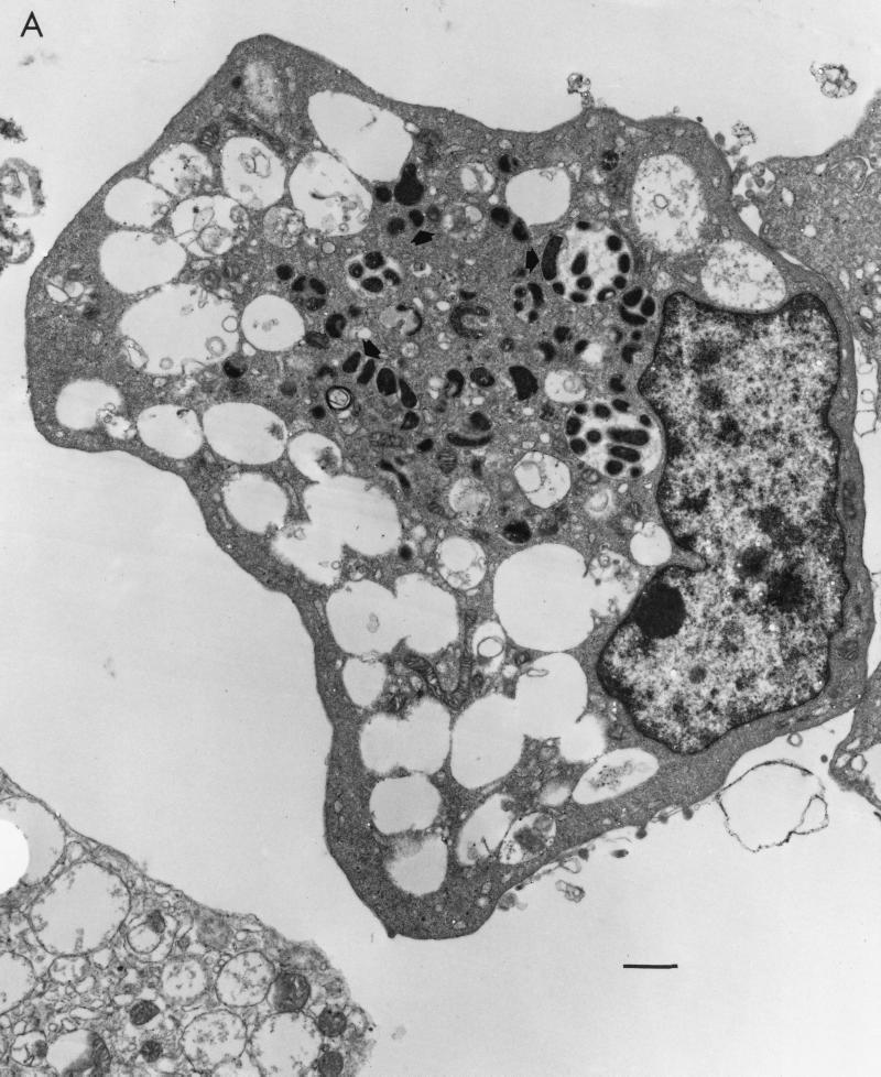
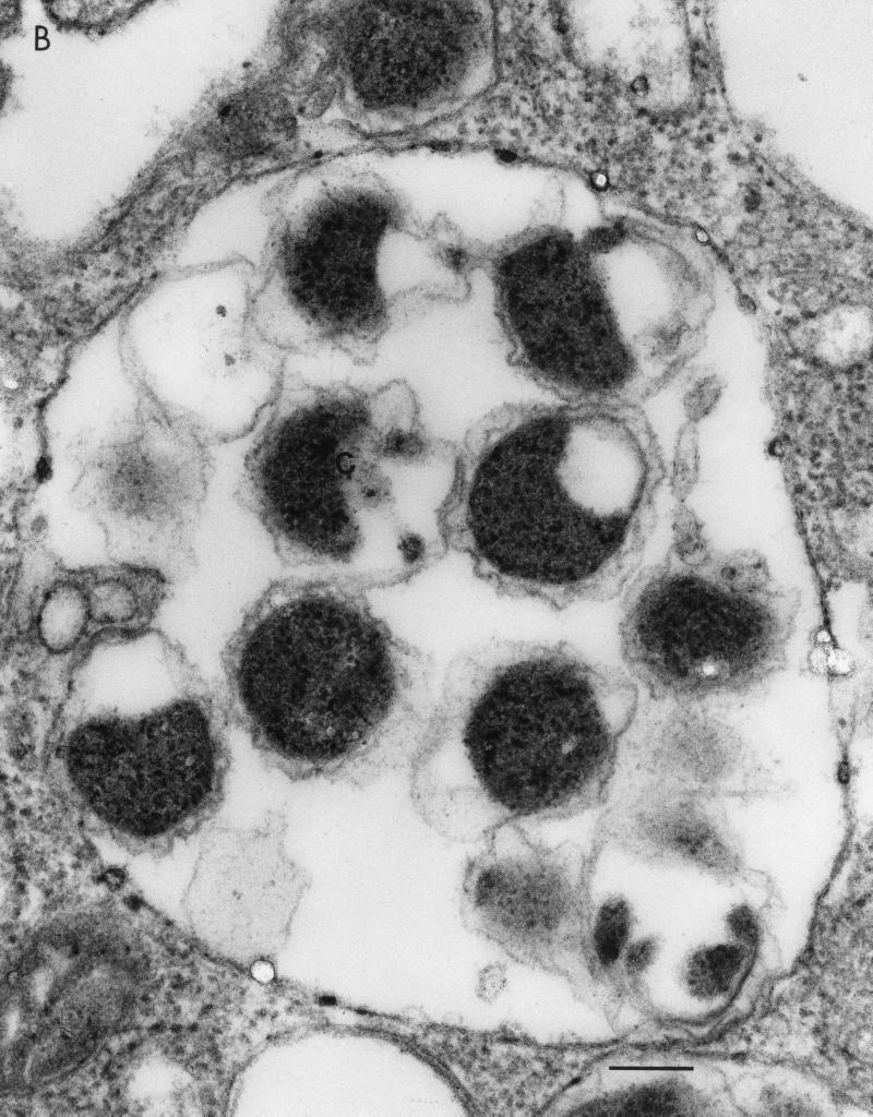
FIG. 1.
(A) Transmission electron micrograph of ehrlichial organisms (arrows) in cytoplasmic vacuoles of mouse macrophage culture. Bar, 1 μm. (B) Transmission electron micrograph, at a higher magnification, showing ultrastructure of the culture-derived causative agent of equine monocytic ehrlichiosis. Single organisms are surrounded by a distinct cytoplasmic vacuolar membrane. Note the rippled outer membrane of the organism. Bar, 0.2 μm.


