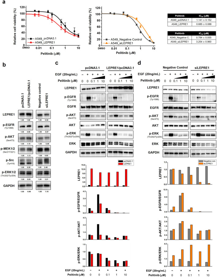
Figure 4.
LEPRE1 regulates drug responsiveness in lung cancer-derived A549 cells. (a) LEPRE1 overexpression (left side) or LEPRE1 knockdown (right side) A549 cells were treated with pelitinib for 3 days. Cell viability was determined using WST-1 proliferation assays. (b) A549 cells were transfected with pcDNA3.1 and LEPRE1/pcDNA3.1 or with Negative control siRNA and LEPRE1 siRNA, as indicated. After 48 h, EGFR, AKT, and ERK expression were determined by western blot analysis. (c,d) A549 cells were transfected with pcDNA3.1 and LEPRE1/pcDNA3.1 (c) or with Negative control siRNA and LEPRE1 siRNA (d) and then treated with EGF and/or the indicated concentration of pelitinib (0, 0.1, 1, 10 μM) for 24 h. Cell extracts were prepared and immunoblotted using the indicated antibodies. GAPDH was used as an internal control. All western blots are pre-cut. Membranes were often stripped and reprobed for multiple antibodies.

