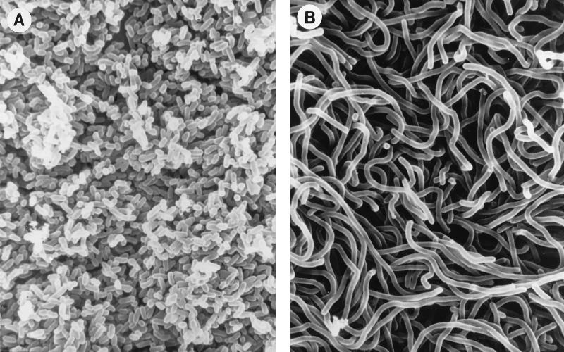
FIG. 1.
Scanning electron micrographs of the short forms seen in colonial type 1 (A) and the filamentous forms seen in colonial type 2 (B) E. rhusiopathiae. A Millipore filter with 72-h growth of E. rhusiopathiae was placed on a 5% sheep blood agar plate. The filter was lifted off and placed in 2% buffered glutaraldehyde. The width of the organism is 0.5 μm.

