FIGURE 5.
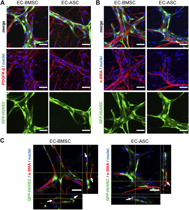
Mesenchymal stem cell transition towards perivascular cells. (A,B) BMSCs as well as ASCs have pericytic characteristics marked by positive expression of PDGFR-β (A) and α-SMA (B). The immunostaining revealed that majority of MSCs are stained for PDGFR-β in both co-cultures. BMSCs are more organized and localize closely to the microvessels whereas ASCs have random appearance scattered throughout the hydrogel. In contrast to PDGFR-β staining, considerably less MSCs were stained for a-SMA in EC-ASC co-culture compared to EC-BMSC co-culture. Scale bars, 50 µm. (C) α-SMA positive MSCs (red) localize in close proximity and wrap around the vascular structures (green) in both EC-BMSC and EC-ASC co-cultures. White arrowheads indicate interaction of pericytes (red) with ECs (green). Scale bars, 50 µm. Nuclei (blue) are stained with DAPI. Donor cell lines BMSC 3 and ASC 1 were used for generation of data presented in (A). Donor cell lines BMSC 1 and ASC 2 were used for generation of data presented in (B, C).

