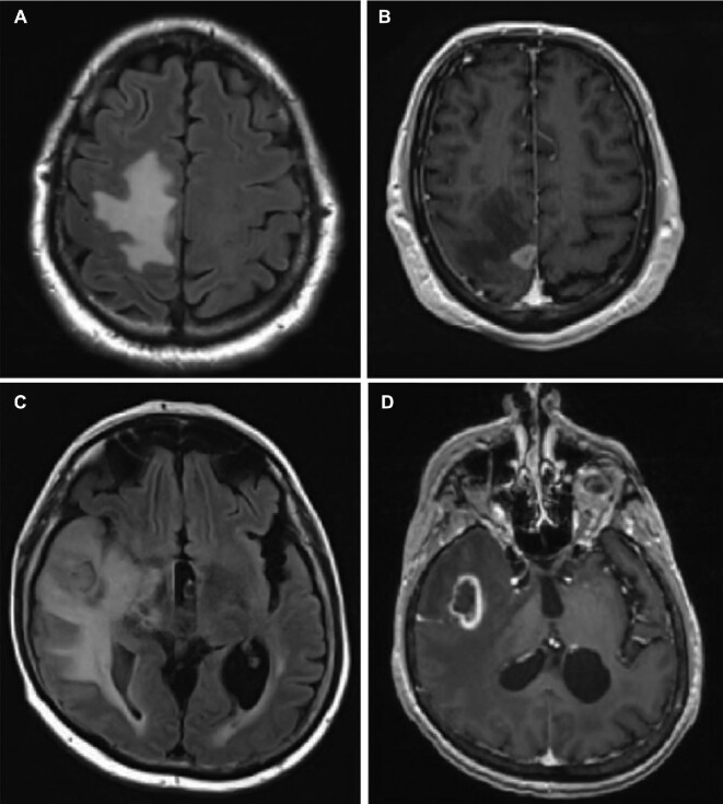
FIGURE 1.
MRIs of representative patients. A, B, Represent recurrent symptomatic brain lesion 6 mo s/p SRS in patient 4 on fluid-attenuated inversion-recovery (FLAIR), A, and T1 weighted MRI + Gd, B. This proved to be recurrent renal cell carcinoma. C, D, Represent recurrent lesion 8 mo s/p SRS in patient 11 on FLAIR and T1 weighted MRI + Gd, which proved to be consistent with RN.

