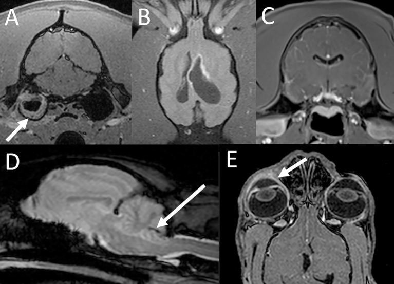
Figure 3.
MRI findings with inflammatory conditions. (A) Otitis media and interna in a tiger. The transverse post contrast T1-W GRE image with FatSat (“VIBE”) shows soft tissue material along the periphery of the right tympanic bulla (arrow). There is also mild contrast enhancement of the soft tissues adjacent to the bulla consistent with regional inflammation. (B) Intracranial blastomycosis in a tiger. On the dorsal T1-W FatSat image there is asymmetric dilatation of the lateral ventricles with marked contrast enhancement of the ventricular lining especially along the rostral horn of the left lateral ventricle. (C) Lymphoplasmacytic meningoencephalitis in a tiger. On the transverse post contrast T1-W GRE image with FatSat (“VIBE”) there is evidence of marked mostly leptomeningeal contrast enhancement. (D) Parasitic meningoencephalitis in a bobcat. A linear susceptibility artifact is associated with the cerebellum (arrow), consistent with a hemorrhagic migration tract. (E) Corneal ulcer, keratitis and uveitis in a lynx. The dorsal post contrast T1-W GRE image with FatSat (“VIBE”) shows contrast enhancement of the right cornea and uvea (arrow) compared to the left.

