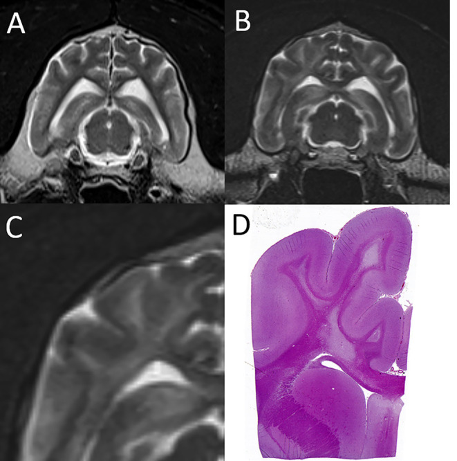
Figure 4.

Leukoencephalopathy in a snow leopard. (A) On this normal transverse T2-W image of the brain in a different leopard, the centrally located finger-like white matter tracts are hypointese to gray matter. (B) In the affected leopard the white matter tracts are diffusely T2 hyperintense. (C,D) Magnification of the T2-W MRI image paired with the corresponding histopathology image, which shows severe pallor and loss of cerebral white matter. Courtesy of Dr. Mee-Ja Sula.
