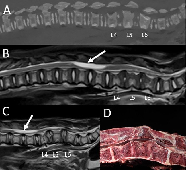
Figure 9.
Vertebral dysplasia in a liger. MRI and CT were performed at 0.75 years of age, and the MRI examination was repeated at 1.25 years of age. (A) Sagittal reconstruction of the CT images, (B) sagittal T2-W image obtained at the first MRI examination, (C) sagittal T2-W image obtained at the repeat MRI examination, and (D) corresponding gross pathology image. Imaging findings include foreshortening and abnormal shape of the L4 through L6 lumbar vertebrae, with decrease in intervertebral disc space width, decrease in size and normal T2 hyperintense signal of the intervertebral discs, dorsal protrusion of the intervertebral discs, lumbar kyphosis, and spinal cord compression. A large cyst-like intramedullary lesion was identified, most consistent with a syrinx with other cystic conditions not excluded (arrow). Autopsy (D) confirmed vertebral dysplasia with severe syringohydromyelia.

