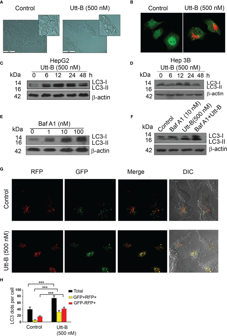
Figure 1.
Utt-B induces autophagy, in vitro. (A) Utt-B treatment induces the formation of vacuolated structures in HepG2 cells. (B) Acidic vacuoles are stained with Acridine Orange (C) Western blot results indicate that LC3II expression is induced in HepG2 cells from 6h to 12h in HepG2 cells. (D) Western blot results indicate that LC3II expression is induced in HepG2 cells from 12h to 24h in Hep3B cells. (E) 10 nM Baf A1 is enough to block autophagy in HepG2 cells, as inferred from LC3-II accumulation. (F) Co-treatment of Utt-B and Baf A1 increases LC3-II accumulation. (G, H) Accumulation of LC3-II by Utt-B was quantitated by RFP-GFP-LC3 tagged protein assay. *** level of significance 3.

