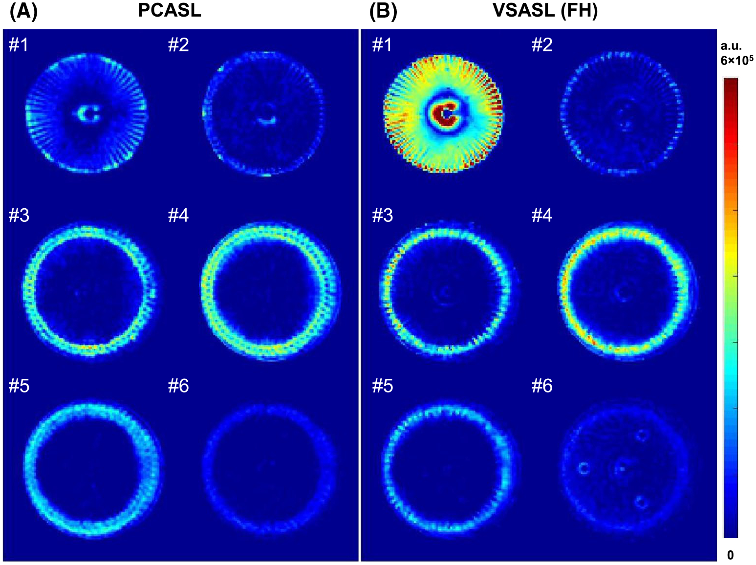
FIGURE 3.

The difference images (subtraction of label and control) of pseudo-continuous arterial spin labeling (PCASL) (A) and velocity-selective arterial spin labeling (VSASL) (B) at the six slices marked by the yellow lines on the sagittal maximum intensity projection (MIP) of VSMRA in Supporting Information Figure S1B at the 350-mL/min flow rate. The VSASL (FH) technique is a velocity-selective inversion-prepared ASL with a foot–head encoding direction. All images are displayed at the same scale
