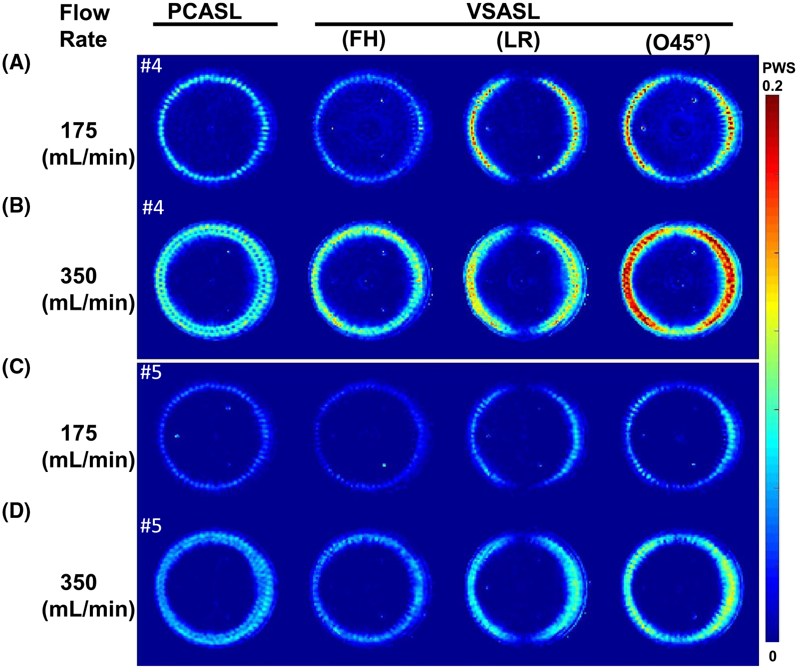
FIGURE 4.

The perfusion-weighted signal (PWS: difference signal normalized by the SIPD) of PCASL and VSASL using FH, LR, and O45°-encoded directions at slice 4 (A,B) and slice 5 (C,D) for 175-mL/min (A,C) and 350-mL/min (B,D) flow rates, respectively
