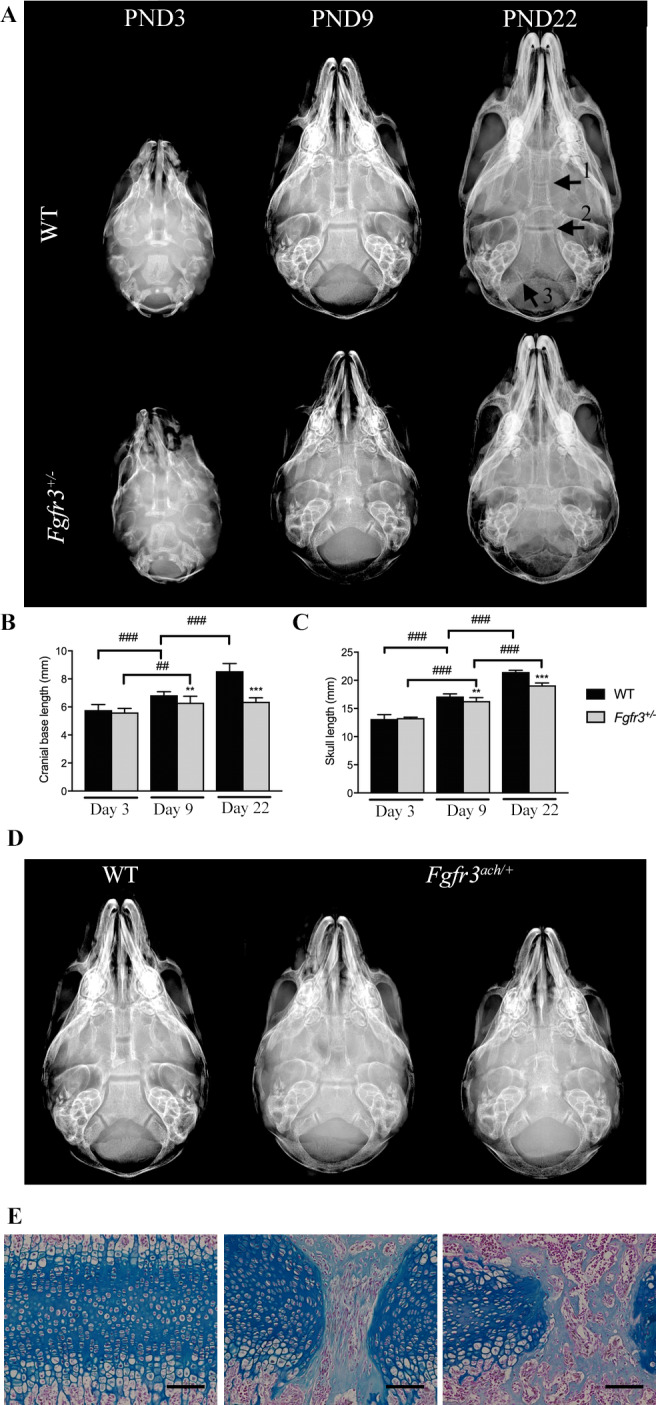
Fig 1.

Premature closure of the SOS is associated with cranial base growth arrest in Fgfr3 ach/+ mice. (A) Representative radiological images of the skull of 3 (PND3), 9 (PND9), 22 (PND22) days old Fgfr3 ach/+ and WT mice. The different cranial base synchondroses are shown with numbered arrows (1: intersphenoid synchondroses [ISS]; 2: spheno‐occipital synchondrosis [SOS]; 3: intraoccipital synchondroses [IOS]). (B, C) Measurements of the cranial base length of 3, 9 or 22 days old WT and Fgfr3 ach/+ mice. (D) Whole skull ventral images of 9 days old WT and Fgfr3 ach/+ mice (E) Alcian blue staining of the SOS of 9 days old WT (left panel) and Fgfr3 +/− mice (middle and right panels), ×10 magnification. ## p < 0.005, ### p < 0.001 compared to matching age and phenotype mice, * p < 0.05, *** p < 0.001 compared to Fgfr3 ach/+ vehicle mice.
