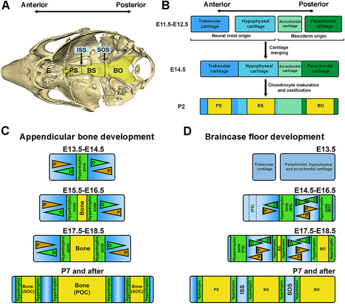
Fig. 1.

A cascade model of braincase floor elongation summarized from studies in this report. (A) A mouse skull model indicates the anatomic location of the braincase floor. ISS = the intersphenoid synchondrosis; SOS = the spheno‐occipital synchondrosis; PS = presphenoid bone; BS = basisphenoid bone; BO = basioccipital bone. (B) Braincase floor development described in previous studies is summarized. Continuous braincase floor is the result of fusion of four cartilages, the parachordal, acrochordal, hypophyseal, and trabecular cartilage from the posterior to the anterior. Then hypertrophic differentiation and subsequent mannerization within parachordal, hypophyseal, and trabecular cartilage leads to formation of BO, BS, and PS in the braincase floor. (C) A model describes appendicular bone elongation. Initial hypertrophy occurs at the middle of cartilage primordia. The growth plate flanking the hypertrophic zone leads to bidirectional elongation of the appendicular bone. (D) A hypothetical model of how the braincase floor elongates. Chondrocytes located at the most posterior end of the braincase floor cartilage primordia start to differentiate to form the hypertrophic zone due to an activity of the remnant notochord, which eventually give rise to the BO. Hypertrophic chondrocytes in this zone subsequently induce differentiation of chondrocytes at the middle of the braincase floor cartilage primordia to form the second hypertrophic zone, which is the future BS, and finally, hypertrophy of the chondrocyte zone for the future PS occurs at the anterior end of the braincase floor cartilage primordia. Three hypertrophic zones will be mineralized when embryogenesis progresses from posterior to anterior, and remaining cartilage will form two mirror‐imaged growth plates, the ISS between PS and BS and the SOS between BS and BO. These synchondroses support the bidirectional growth of each bone, which contributes to the elongation of the braincase floor. D = differentiation; P = proliferation.
