Fig. 6.

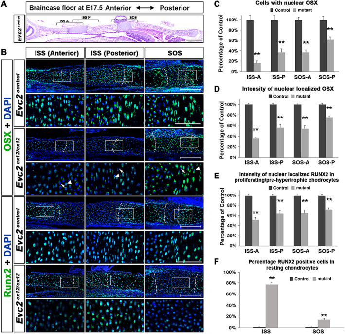
Prehypertrophic differentiation is affected more in the anterior end than the posterior end of the braincase floor. (A) A diagram of the braincase floor in the immunohistochemical studies. (B) Immunodetection of OSX and RUNX2 in the three areas shown in A. The white boxed region was enlarged and shown. Arrows indicate Evc2 mutant chondrocytes with no nuclear localized OSX; arrowheads indicate Evc2 mutant chondrocytes with decreased nuclear localized OSX. Scale bar = 200 μm. Dashed lines indicate the boundary between resting and proliferating chondrocytes. (C) Number of cells with nuclear localized OSX are quantified and shown as percentage of controls, n = 3, **p < 0.01. (D) The intensity of nuclear localized OSX was quantified and shown as a percentage of controls, n = 4, **p < 0.01, error bars denote standard deviations. (E) The intensity of immunosignals for nuclear RUNX2 was quantified and shown as a percentage of control, n = 4, **p < 0.01, error bars denote standard deviations. (F) The percentages of resting chondrocytes with nuclear localized RUNX2 were quantified and shown, n = 3, **p < 0.01. Scale bar = 200 μm, bar in enlarged picture = 20 μm.
