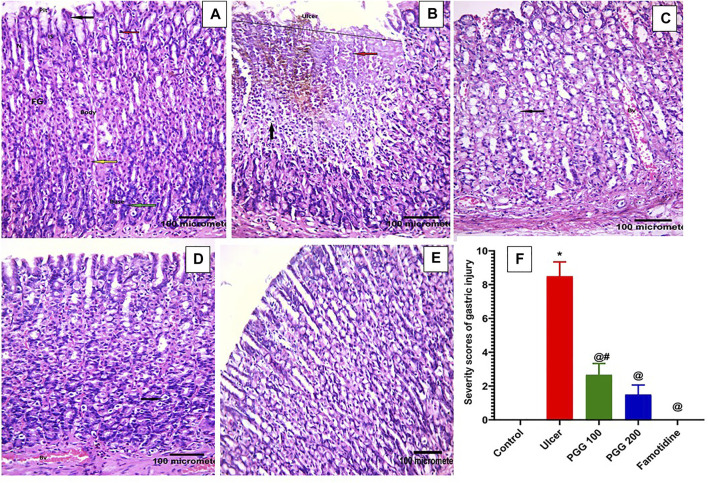
FIGURE 4.
Histopathological effects of pentagalloyl glucose (PGG) effects on the stomach mucosal tissues of animals. Representative gastric sections from (A) normal control group, (B) ulcerative animals (indomethacin alone, 60 mg/kg), (C) indomethacin + PGG-treated groups (100 mg/kg), (D) indomethacin + PGG-treated groups (200 mg/kg), and (E) indomethacin + famotidine-treated group (10 mg/kg). (F) Column graph illustrates the severity scores of gastric injuries among groups, Data are presented as mean ± SEM, *,@,# p < 0.05 vs. control, ulcer, and famotidine groups, respectively, by ANOVA followed by Dunnett’s test. Fundic glands (FGs), gastric pits (Pits), isthmus (IS), neck (N), columnar epithelial cells (black arrow), mucous neck cells (red arrow), pyramidal oxyntic cells (yellow arrow), peptic cells or vacuolation (green arrow), tissue erosion and degeneration (black arrow), exfoliated cells (red arrow), extravasated blood (orang arrow), cellular infiltration (circle), and congested blood vessels (BV), [H&E × 200].

