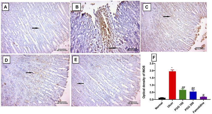
FIGURE 6.
Immunohistochemical staining of inducible nitric oxide synthase (iNOS) in gastric mucosal tissues. (A) Control and (E) famotidine-pretreated ulcer groups furnished a minimal positive cytoplasmic immunoreactivity to iNOS in the gastric mucosal cells (arrows). (B) Ulcerative animals (indomethacin group) displayed an intense positive immune reaction to iNOS in the gastric mucosal cells (arrows). (C,D) PGG -pretreated ulcer groups (100 or 200 mg/kg) showed a reduction in iNOS immunoreactivity (arrows). iNOS immunostaining (avidine biotin peroxidase stained with Hx counter stain X 200, scale bar = 100 µm). (F) Quantitative analysis of immunoreactivity intensity of iNOS. Results are shown as mean ± SEM, n = 10. * @, #significantly different compared the control group, indomethacin, and famotidine groups, respectively, at p < 0.05 by one-way ANOVA followed by Tukey’s post hoc test.

