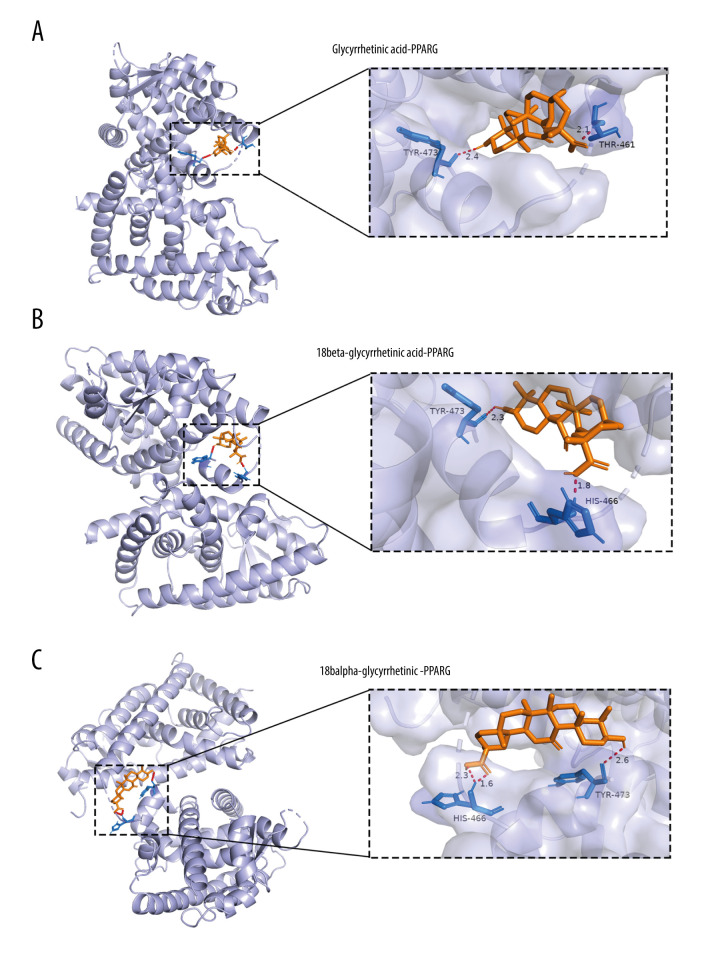
Figure 7. Molecular docking models of glycyrrhetinic acid (A), 18beta-glycyrrhetinic acid (B), and 18alpha-glycyrrhetinic (C) binding to PPARG by AutoDock 4.2 software.
Purple, orange, red, and blue represent protein receptor, small drug ligand, hydrogen bond, and amino acid residue, respectively. The length of bond is added to the bond.

