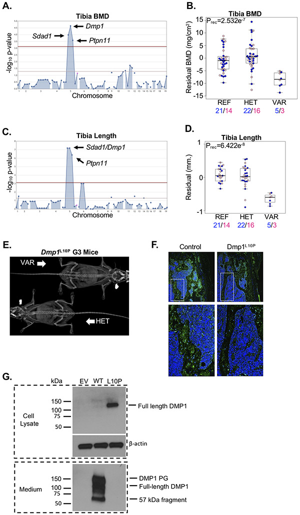
Figure 6.
Loss of DMP1 secretion does not cause severe chondrodysplasia. (A) A single locus segregating alleles in Sdad1, Dmp1, and Ptpn11 were significantly associated with variation in tibia BMD. (B) Mice homozygous for the Dmp1L10P allele (VAR) developed reduced tibia BMD compared to mice heterozygous for the allele (HET) and mice homozygous for the reference allele (REF). Male and female mice are shown with blue and pink symbols, respectively, and the numbers of mice are shown below. Data are median with interquartile range (IQR); whiskers extend up to 1.5x the IQR. (C) A single locus segregating alleles in Sdad1, Dmp1, and Ptpn11 were significantly associated with variation in tibia length. (D) Mice homozygous for the Dmp1L10P allele (VAR) developed slightly reduced tibia length compared to mice heterozygous for the allele (HET) and mice homozygous for the reference allele (REF). Male and female mice are shown with blue and pink symbols, respectively, and the numbers of mice are shown below. Data are median with interquartile range (IQR); whiskers extend up to 1.5x the IQR. (E) Representative radiograph of a mouse homozygous (VAR) or heterozygous (HET) for the Dmp1L10P allele. (F) Immunofluorescence localization of DMP1 protein (green) in the distal femur of a mouse homozygous for the reference allele (Control) or homozygous for the Dmp1L10P allele. DMP1 protein is primarily restricted to the nucleus with little evidence of secretion in the homozygous Dmp1L10P mouse. (G) Western blot analysis of MC3T3-E1 cells transiently expression HA-tagged wild-type DMP1 (WT) or mutant DMP1L10P (L10P). Cell lysate and medium were analyzed separately to detect intracellular and secreted DMP1 protein, respectively. The DMP1 proteoglycan (DMP1 PG) and 57kDa fragment are evident in the culture medium. Beta-actin is shown as a loading control. Empty vector (EV) is shown as negative control.

