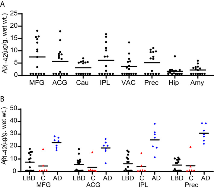
Figure 4.

Regional distribution of insoluble Aβ(1‐42) concentration in LBD, Control, and AD cases. Levels of insoluble Aβ(1‐42) were measured by sandwich ELISA. (A) Eight of the 15 cases had measurable Aβ levels in most brain regions. For LBD cases with measurable Aβ, the highest levels of Aβ were found in MFG, ACG, IPL, and Precuneus. (B) Aβ levels were measurable in two of the six control (red triangles) cases. One control case had high levels of Aβ in all brain regions. AD (blue diamonds) cases had significantly higher Aβ levels compared with the subgroup of LBD (black circles) cases that had detectable Aβ levels in all four brain regions examined. The LLQ for the ELISAs was 14.17 pg/mL or 0.63 µg/g wet wt tissue. Data were analyzed with the two‐tailed Mann–Whitney test using a significance level of 0.05 and corrected for multiple comparisons with the Holm‐Bonferroni method. 52
