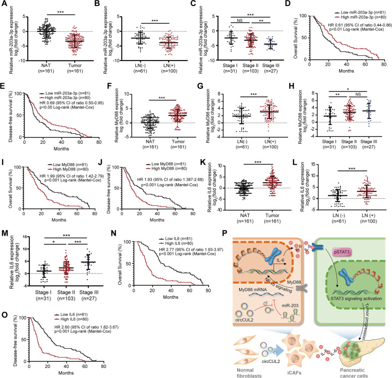
Fig. 8.
Clinical implication of circCUL2/miR-203a-3p/IL6 axis in PDAC. A, F, K The relative expression of miR-203a-3p (A), MyD88 (F) and IL6 (G) in 161 PDAC tissues compared to paired NAT. B-C, G-H, L-M Association analysis between miR-203a-3p (B-C), MyD88 (G-H) and IL6 (L-M) expression levels and LN status and tumor stages in 161 PDAC tissues. (D-E, I-J, N-O) Kaplan-Meier analysis of the correlation between miR-203a-3p (D-E), MyD88 (I-J) and IL6 (N-O) expression levels and OS or DFS of 161 PDAC patients. The median miR-203a-3p, MyD88 and IL6 expression was used as the cutoff value. P Proposed model indicates the mechanism by which circCUL2 activated iCAF phenotype and production of IL6 to promote malignant progression of PDAC via miR-203a-3p/MyD88/NF-κB pathway. Data are expressed as the mean ± SD. *p < 0.05 and ***p < 0.001

