Fig. 5.
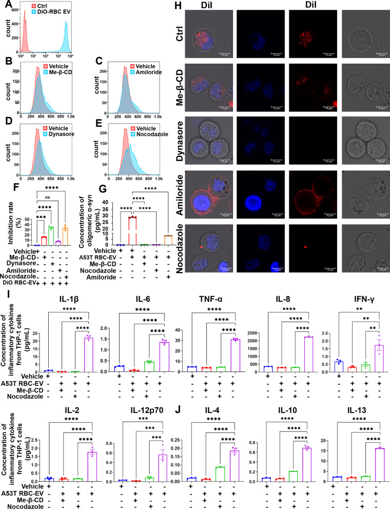
Endocytosis was involved in immune sensitization of monocytes induced by A53T RBC-EVs. A RBC-EVs uptake by THP-1 cells was determined by flow cytometry. Red histogram represented THP-1 cells without RBC-EVs, while blue histogram represented THP-1 cells containing RBC-EVs. The RBC-EVs were labelled with DiO. B–E Effects of endocytosis inhibitors on RBC-EVs uptake in THP-1 cells was determined by flow cytometry. Red histogram represented THP-1 cells co-treated with RBC-EVs and vehicle control, while blue histogram represented THP-1 cells co-treated with RBC-EVs and endocytosis inhibitor. F Quantitative analysis of inhibition ratio of Me-β-CD, Dynasore, Amiloride and Nocodazole. G Levels of oligomeric α-syn in THP-1 cells pretreated with A53T RBC-EVs, along with Me-β-CD, Nocodazole or Amiloride. H Representative confocal image of THP-1 cells incubated with DiI (red)-labelled RBC-EVs after treatment with Dynasore, Nocodazole Me-β-CD and Amiloride, cell nucleus was labelled by Hoechst 33258. Scale bar, 10 µm. I Quantitative analysis of pro-inflammatory cytokines IL-1β, IL-6, TNF-α, IL-8, IFN-γ, IL-2, and IL-12p70, and J anti-inflammatory cytokines IL-4, IL-10, and IL-13 using MSD, released by THP-1 cells pretreated with A53T RBC-EVs alone or with Nocodazole or Me-β-CD, followed by LPS stimulation. N = 3 independent experiments in each group. Values are means ± S.E.M, one-way ANOVA test. *, P < 0.05; **, P < 0.01; ***, P < 0.001; ****, P < 0.0001

