Fig. 3—


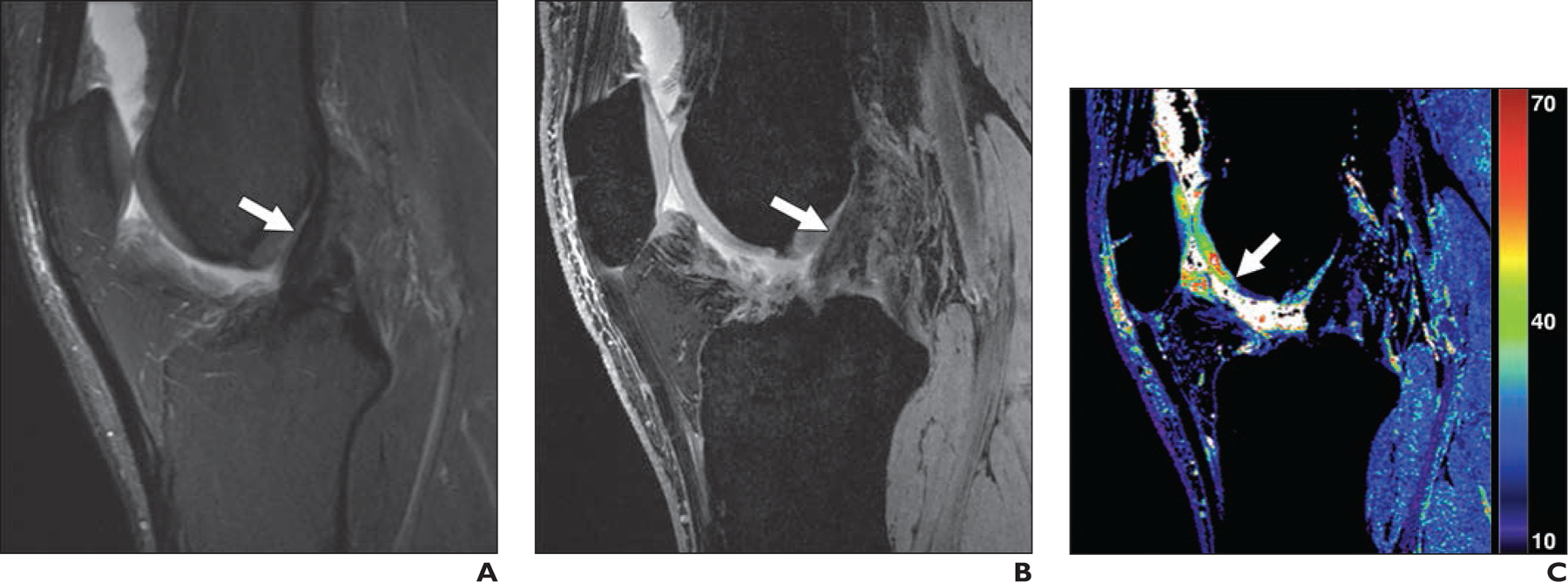
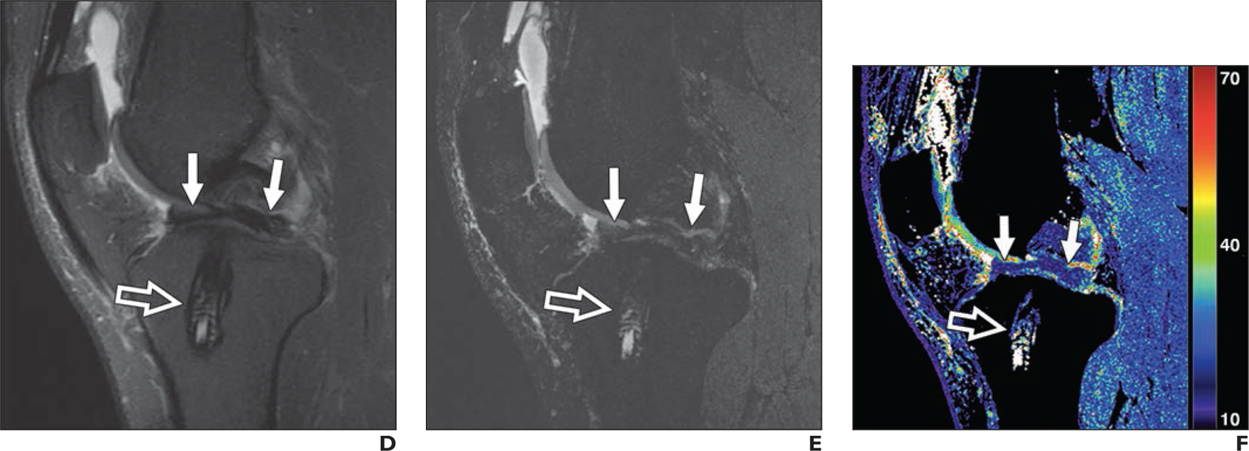
30-year-old man with previous anterior cruciate ligament (ACL) reconstruction who experienced knee reinjury.
A, Sagittal T2-weighted MR image shows intact ACL graft with conspicuity (arrow).
B, Quantitative double-echo steady-state (qDESS) MR image with mixed T2- and T1-weighted echo shows conspicuity of ACL graft (arrow) similar to A.
C, qDESS T2 map (values in milliseconds) shows focal abnormal T2 relaxation (arrow) despite cartilage in trochlea graded as normal in A and B.
D, Sagittal T2-weighted MR image shows biointerference screw in tibial tunnel (open arrow) and arthroscopically proven bucket-handle tear of medial meniscus (solid arrows) with fragment displaced in intercondylar notch.
E and F, qDESS image with higher T2-weighted echo (E) and qDESS T2 map (F) show biointerference screw (open arrow) and meniscal tear (solid arrows) similar to D.
