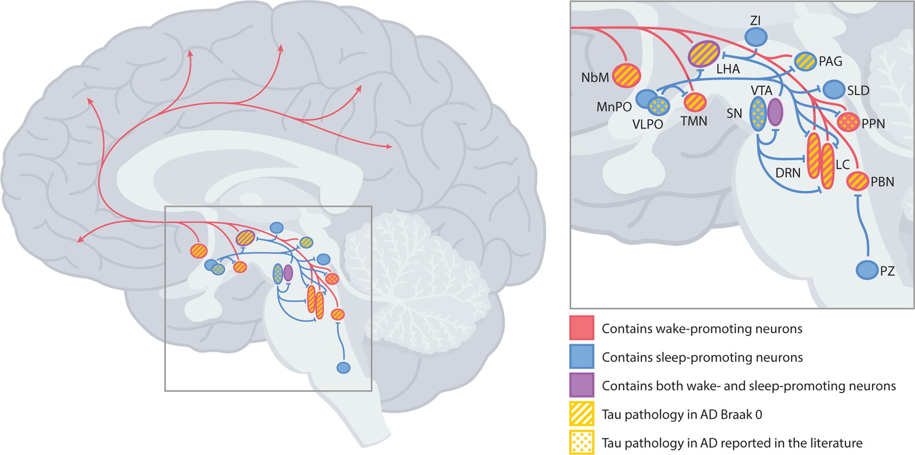
Fig. 2.

Brain regions in the brainstem, hypothalamus, and basal forebrain that contain sleep- and wake-promoting neurons, with areas containing abnormal tau and/or neurofibrillary tangles in AD noted. Red arrows indicate wake-promoting projections. Blue lines indicate sleep-promoting projections. DRN = dorsal raphe nucleus; LC = locus coeruleus; LHA = lateral hypothalamic area; MnPO = median preoptic nucleus; NbM = nucleus basalis of Meynert; PAG = periaqueductal gray; PBN = parabrachial nucleus; PPN = pedunculopontine nucleus; PZ = parafacial zone; SLD = sublaterodorsal nucleus; SN = substantia nigra; TMN = tuberomammillary nucleus; VLPO = ventrolateral preoptic nucleus; VTA = ventral tegmental area; ZI = zona incerta. (For interpretation of the references to color in this figure legend, the reader is referred to the Web version of this article.)
