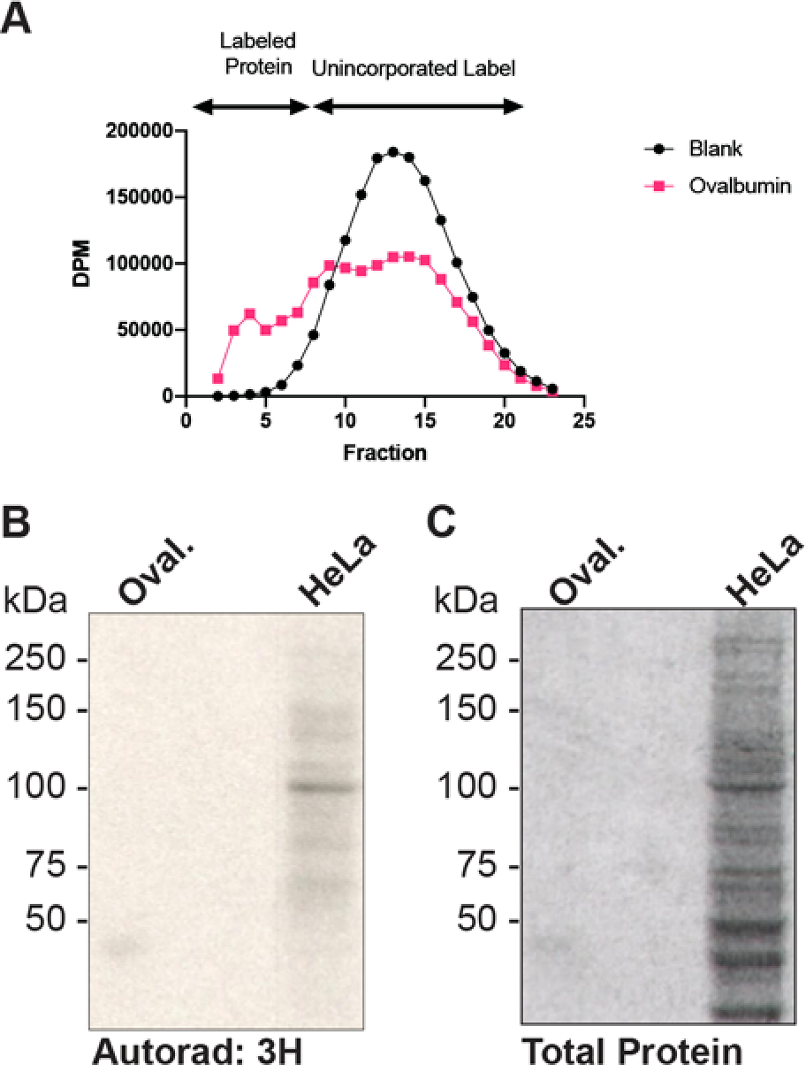
Figure 5. Galactosyltransferase Labeling.

A) Ovalbumin (•, pink) or a blank (■, black) were labeled with Gal-T and 3H-UDP-Gal. Reactions were desalted using a G50 column. Fractions were counted and those with significant signal above the blank were combined and acetone precipitated. B) Ovalbumin (51,722 DPM) and HeLa cell lysates (18,295 DPM) labeled with Gal-T were separated by SDS-PAGE and stained with Coomassie blue (total protein). The resulting gel was dried and exposed to autoradiography film (1 week) with the aid of an intensifying screen.
