Figure 4.
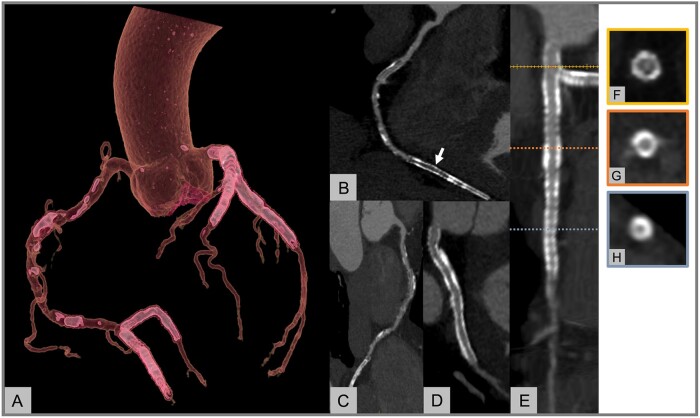
CCTA in patients with multiple coronary stents. (A) A 58-year-old man underwent CCTA due to recent onset of atypical chest pain. The patient had prior multiple stenting as shown in the volume rendering reconstruction of the coronary tree. (B–D) Curved MPRs of RCA-PDA (B), RCA-PL (C) and LCX (D). The stent lumen on the PDA artery (B, arrow) appears homogenously hypodense indicating stent occlusion. (E–H) Straight MPR of LM-LAD (E) and cross-sectional images of distal LM (F) as well as proximal (G) and distal (H) LAD. CCTA demonstrated a dark rim in the distal LM stent documenting the presence of in-stent restenosis (F). While the stent in the proximal LAD (G) was assessable and judged as patent, the small size of the stent in the distal LAD (H) precluded the evaluation of the lumen. CCTA, coronary computed tomography angiography; LAD, left anterior descending Artery; LCX, left circumflex artery; LM, left main; MPR, multiplanar reconstruction; PDA, posterior descending artery; PL, posterolateral branch; RCA, right coronary artery.

