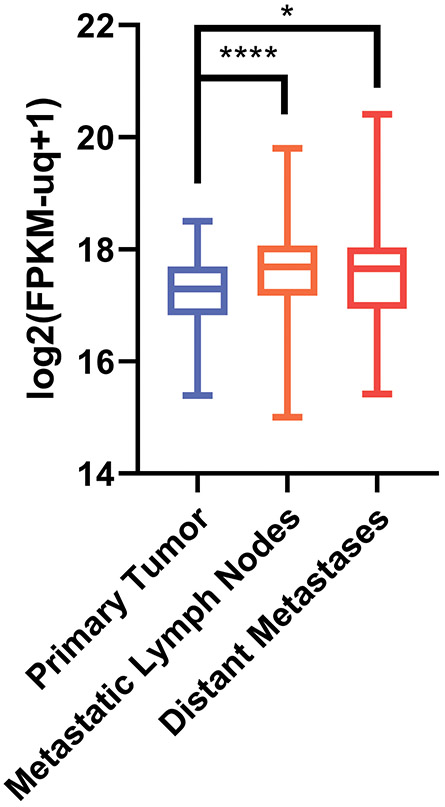
Figure 1. PHF10 mRNA expression (RSEM counts) in cutaneous melanoma.
In GDC TCGA Melanoma cohort (n=477) primary tumors exhibited a lower expression of PHF10 compared to metastatic lymph nodes and distant sites (Kruskal-Wallis test with post hoc Mann Whitney U test and Benjamini-Hochberg correction for multiple comparisons was used to assess the significance). TCGA cutaneous melanoma RNA-Seq dataset (https://cancergenome.nih.gov/) revealed a statistically significant increase of PHF10 transcripts in metastatic vs primary melanoma specimens (*p<0.01, ****p<0.00001). Y axis: log2 FPKM (fragments/kilobase of exon model/million reads mapped).

