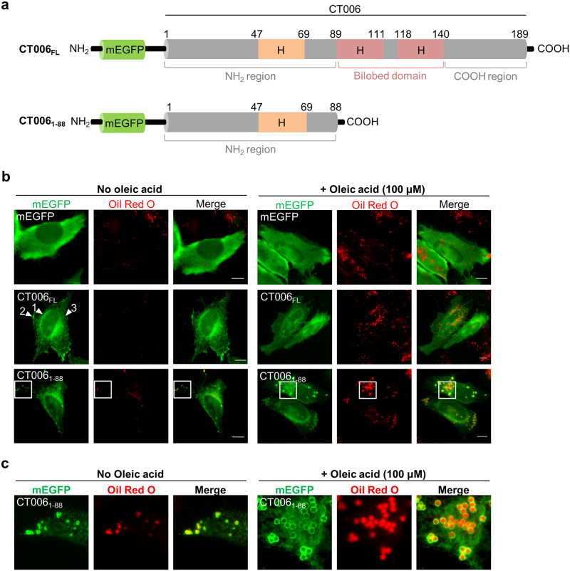
Fig 3. The first 88 amino acid residues of CT006 fused to mEGFP co-localize with LDs in mammalian cells.
HeLa 229 cells were transfected with plasmids encoding mEGFP or different regions of CT006 containing a mEGFP tag at their amino-termini (mEGFP-CT006 proteins). (a) Schematic representation of mEGFP-CT006 and mEGFP-CT0061-88 (not drawn to scale); H, Putative hydrophobic domain. (b) At 18 h post-transfection, cells were either treated with ethanol (solvent control; left-hand side panel) or 100 μM oleic acid (right-hand side panel) for 6 h and then fixed with 4% (w/v) PFA. Fixed cells were labeled with anti-GFP and the appropriate fluorophore-conjugated secondary antibody, stained with Oil Red O (3:2 v/v Oil Red O stock solution diluted in water), and imaged by fluorescence microscopy. Scale bars, 10 μm. Arrowheads indicate the reticular distribution of mEGFP-CT006FL (1) and the accumulation in patches near the plasma membrane (2) or cytosol (3). (c) In the area delimited by white squares (Fig 3b) images were zoomed.

