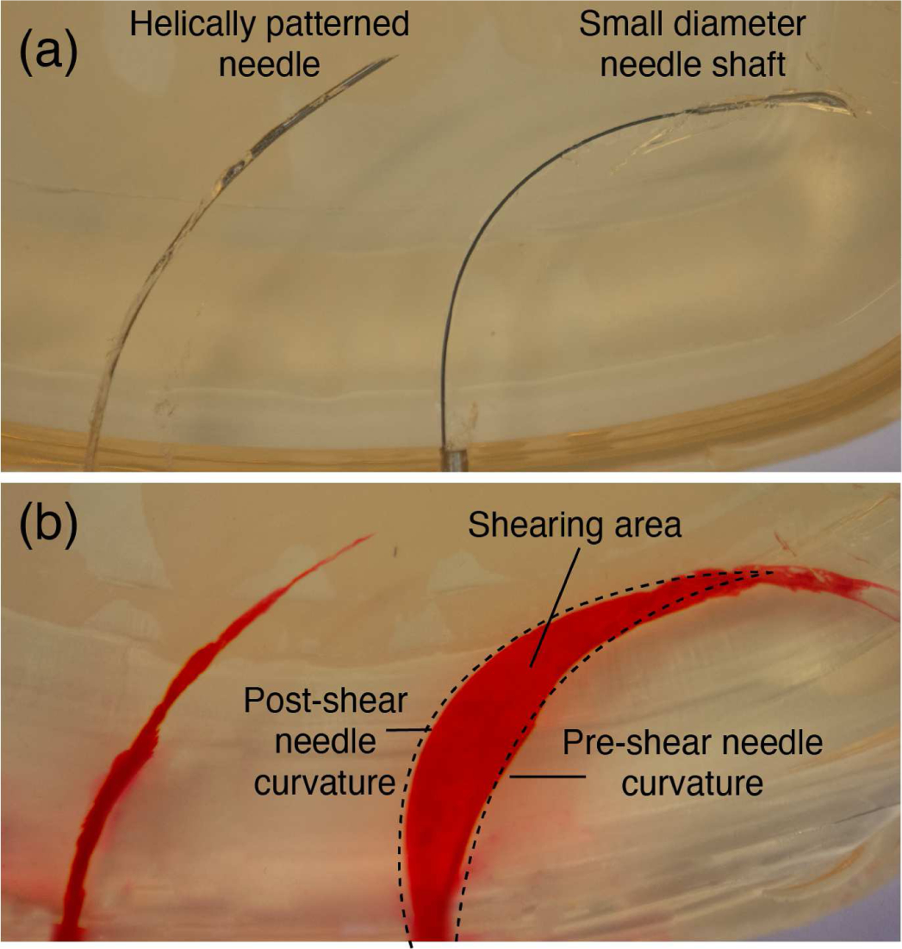
Fig. 4:

Helically patterned needle inserted in tissue vs. a needle with a small diameter shaft (0.36 mm OD, 0.24 mm ID) with a large bent tip. (a) shows the patterned needle (left) and the small shaft needle (right) inserted in the gelatin. (b) shows the tissue damage caused by each insertion. Red dye was injected into the path created by each needle after the needle was removed from the gelatin.
