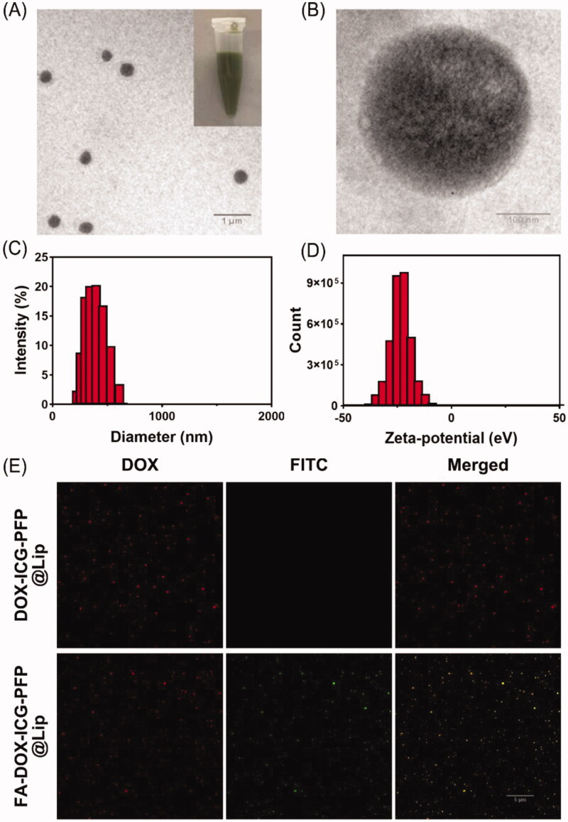
Figure 1.
Morphology and characterization of liposomes. (A) Transmission-electron micrograph of FA-DOX-ICG-PFP@Lip. (Inset) Images of the FA-DOX-ICG-PFP@Lip suspension. (B) High-magnification transmission-electron micrograph of FA-DOX-ICG-PFP@Lip. (C) Size distribution of FA-DOX-ICG-PFP@Lip. (D) Zeta potentials of FA-DOX-ICG-PFP@Lip. (E) The detection of the folate on the surface of FA-DOX-ICG-PFP@Lip by laser scanning confocal microscopy.

