
Fig. 10.
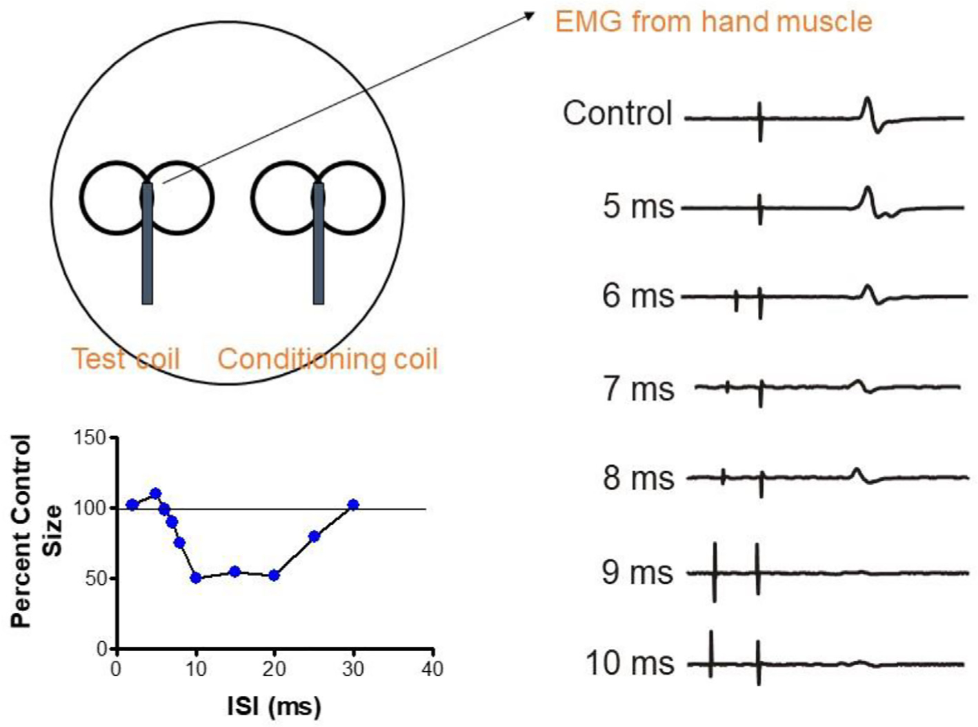
Interhemispheric inhibition between the motor cortices. A suprathreshold TMS pulse (test pulse) is applied to the left hemisphere to evoke a motor evoked potential (MEP) (control trace in right panel). If a conditioning TMS pulse is applied 5–10 ms beforehand, it suppresses the amplitude of the evoked MEP, starting at an interval of 6 ms. The panel in the bottom left shows the time course of inhibition, where the amplitude of the control MEP is set to 100%. The duration and depth of inhibition depend on the intensity of the conditioning stimulus (not shown).