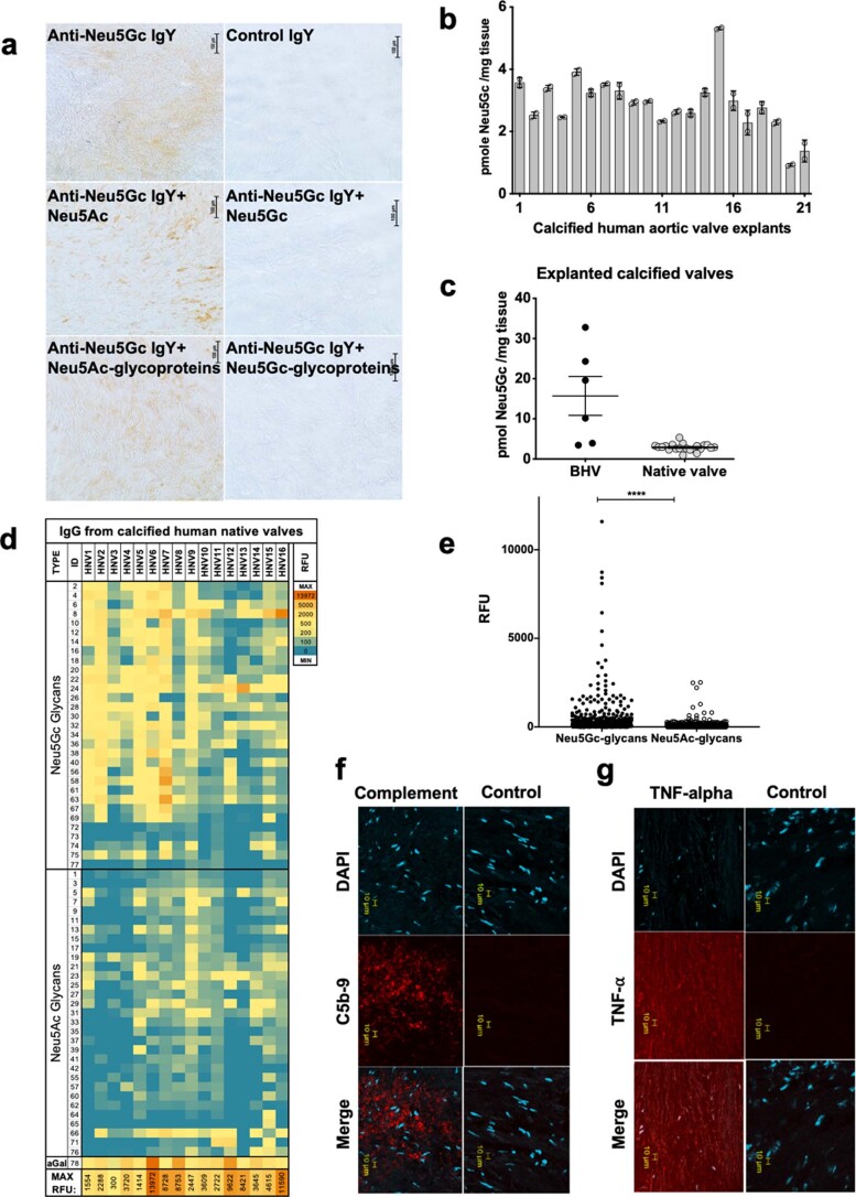
Extended Data Fig. 3. Neu5Gc and anti-Neu5Gc IgG in explanted calcified native aortic human heart valves.
a, Immunohistochemistry of explanted calcified native human aortic heart valve tissues revealed specific detection of Neu5Gc, by staining with chicken anti-Neu5Gc IgY versus control chicken IgY. Neu5Gc staining was inhibited by precomplexing of primary antibody with free Neu5Gc or Neu5Gc-glycoproetins, but not with Neu5Ac or Neu5Ac-glycoproetins (representative of at least two independent experiments). b, Quantitative analysis of Neu5Gc in explanted calcified native aortic valves by DMB-HPLC (n = 21 explants; mean±sem; two independent experiments). c, Neu5Gc DMB-HPLC quantification shows much higher Neu5Gc levels in explanted calcified BHVs compared to explanted calcified native valves (mean±sem; n = 6, 21 BHV and native valve explants, respectively). d, IgG antibodies were purified from homogenized calcified native valve explants (n = 16) by protein A, then analyzed by glycan microarray printed with αGal and Neu5Gc/Neu5Ac-glycans and relative fluorescence units (RFU) determined after scanning, showing anti-αGal IgG and anti-Neu5Gc IgG response in all analyzed samples. e, Glycan microarrays analysis of IgG purified from explants (n = 16) revealed strong recognition of Neu5Gc-glycans while minimal recognition of Neu5Ac-glycans (mean±sem; each spot is IgG binding to a specific glycan on the array of all BHV samples analyzed; Two-tailed Mann-Whitney test, ****, p < 0.0001). f, Immunohistochemistry of explanted calcified native valves with mouse-anti-human-C5b-9 IgG shows staining of membrane attack complex (representative of three independent experiments). g, Immunohistochemistry of explanted calcified native valves with biotinylated-rabbit-anti-human TNF-α, detected with Cy3-streptavidin, shows deposition of TNF-α (n = 1).

