Fig. 6.
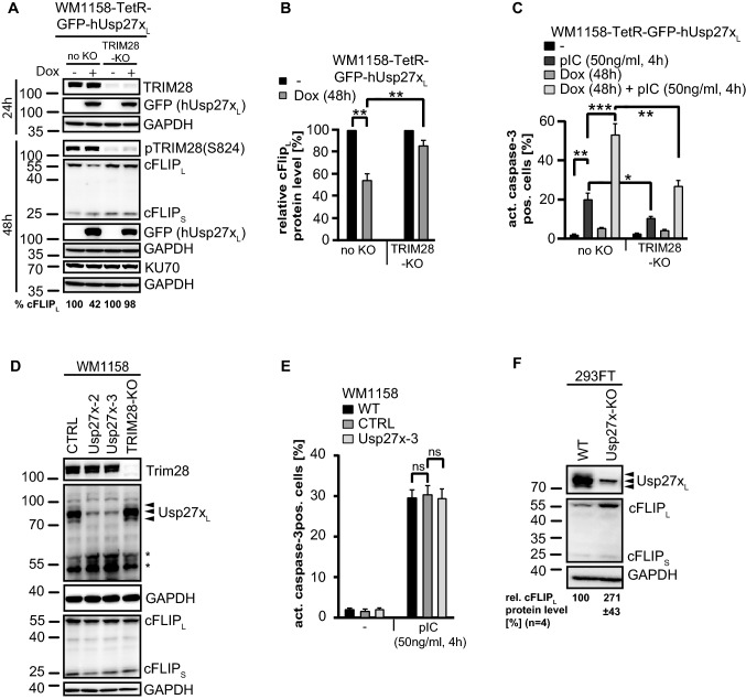
Loss of cFLIPL by Usp27xL expression requires TRIM28, which is required for pIC induced apoptosis and deficiency of Usp27xL stabilizes cFLIPL in 293FT cells. A, B, hUsp27xL induction decreases cFLIPL protein levels in a TRIM28 dependent manner. GFP-hUsp27xL doxycycline-inducible WM1158 (no KO) or same cells where the TRIM28 locus had been targeted by CRISPR/Cas9 to generate TRIM28 deficient cells (TRIM28-KO) were stimulated as indicated, lysed in Laemmli buffer and protein levels were determined by Western blot (n = 4). B, Quantification of cFLIPL protein levels detected by Western blotting (A) after 48 h dox treatment normalized to GAPDH. Shown are the cFLIPL protein levels compared to the untreated respective cell line (**: p value < 0.005, error bars represent SEM; n = 4). C, TRIM28 is needed for pIC induced cell death. Same cells as in (A) were stimulated as indicated, harvested, fixed and stained for active caspase-3 followed by FACS analyses. Data (means, n = 7; error bars represent SEM). *, **, ***: significant, see 2 section. D, Usp27x- or TRIM28 deficiency does not change endogenous cFLIP level and TRIM28 deficiency does not change Usp27x level (and vice versa) in WM1158 cells. Whole-cell-lysates from control (CTRL), polyclonal Usp27x-deficient (Usp27x-2 and Usp27x-3), or TRIM28-deficient (TRIM28-KO) WM1158 melanoma were run on SDS-gels and endogenous level of TRIM28, Usp27x (using custom-made Usp27x antibody) and cFLIP level were detected by Western blotting. Arrow-heads indicate the specific signal for human Usp27xL, asterisks indicate unspecific signal (n = 3). E, Usp27x-deficiency does not protect WM1158 cells from pIC-induced apoptosis. WM1158 cells were stimulated with pIC as indicated and apoptosis was measured as described in C (n = 3, Data (means, ns: not significant)). Error bars represent SEM. F, deficiency of Usp27xL stabilises cFLIPL in 293FT cells. Same 293FT wild-type (WT) or Usp27x-deficient 293FT cells as described in Figs. 1B and 5D were analysed for endogenous cFLIP level (n = 4). Quantification of cFLIPL protein levels detected by Western blotting normalized to GAPDH is indicated. Shown are the relative cFLIPL protein levels (%) compared to the respective WT cell line (p value < 0.05, ± represent SEM; n = 4)

