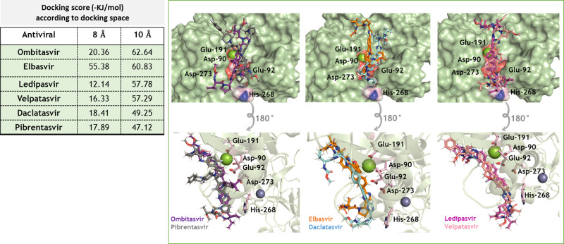
Fig. 2. 3D representation of the best docking poses for HCV NS5A inhibitors in the nsp14 exonuclease active site.
The NS5A inhibitors were built and minimized in terms of energy by density functional theory (DFT). Docking was performed using GOLD 2020.2 software with ChemPLP as scoring function. Because of the diversity in NS5A inhibitors’ molecular weights, they were allowed to dock within 8 or 10 Å spheres in the SARS-CoV-2 nsp14 exonuclease active site, constructed in Supplementary Fig. S-2. The NS5A and catalytic amino acid residues are in stick representation. Mg++ and Zn++ are represented as green and indigo blue spheres, respectively.

