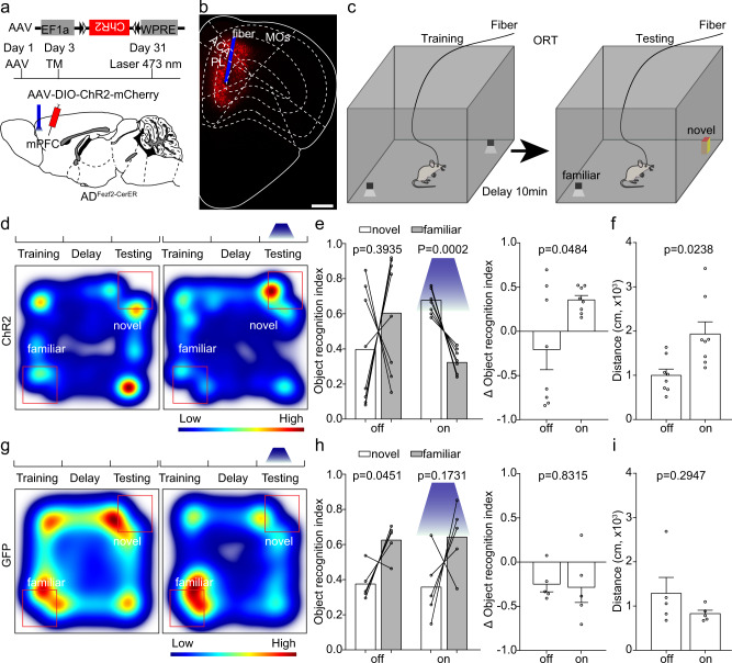
Fig. 2. Activation of extratelencephalic projection neurons in the mPFC of 5×FAD mice improved the object recognition memory expression.
a The experimental strategy of activation of extratelencephalic projection neurons in the mPFC. b An example of position of optical fiber for activation of the extratelencephalic projection neurons in the mPFC. c The experimental procedure of novel object recognition test. d Heat-map plots showed object recognition memory measures from the object recognition test. Red = more time, blue = less time. The heatmap in the left and right showed examples of ADFezf2-CreER mice that expressed ChR2 in the ET neurons in the mPFC exploring the familiar object and novel object during the test session without or with light stimulation, respectively. e Statistical plots of object recognition index and delta object recognition index in laser off and laser on sessions. (left and right, two-tailed paired t test). n = 8 animals for each group. f Statistical plots showed the traveling distance during the test session with or without light stimulation. two-tailed paired t test; n = 8 animals. g The heatmap in the left and right showed examples of ADFezf2-CreER mice that expressed GFP in the ET neurons in the mPFC exploring the familiar object and novel object during the test session without or with light stimulation, respectively. h Statistical plots of object recognition index and delta object recognition index in laser off and laser on sessions. (left and right, two-tailed paired t test). n = 5 animals. i Statistical plot showed that the light stimulation in control group that expressed GFP did not alter the traveling distance during the test. two-tailed paired t test, n = 5 animals. Scale bar in b is 500 μm. All data are listed as the Mean ± SEM. ACA anterior cingulate area, PL prelimbic area, MOs secondary motor area, TM tamoxifen.

