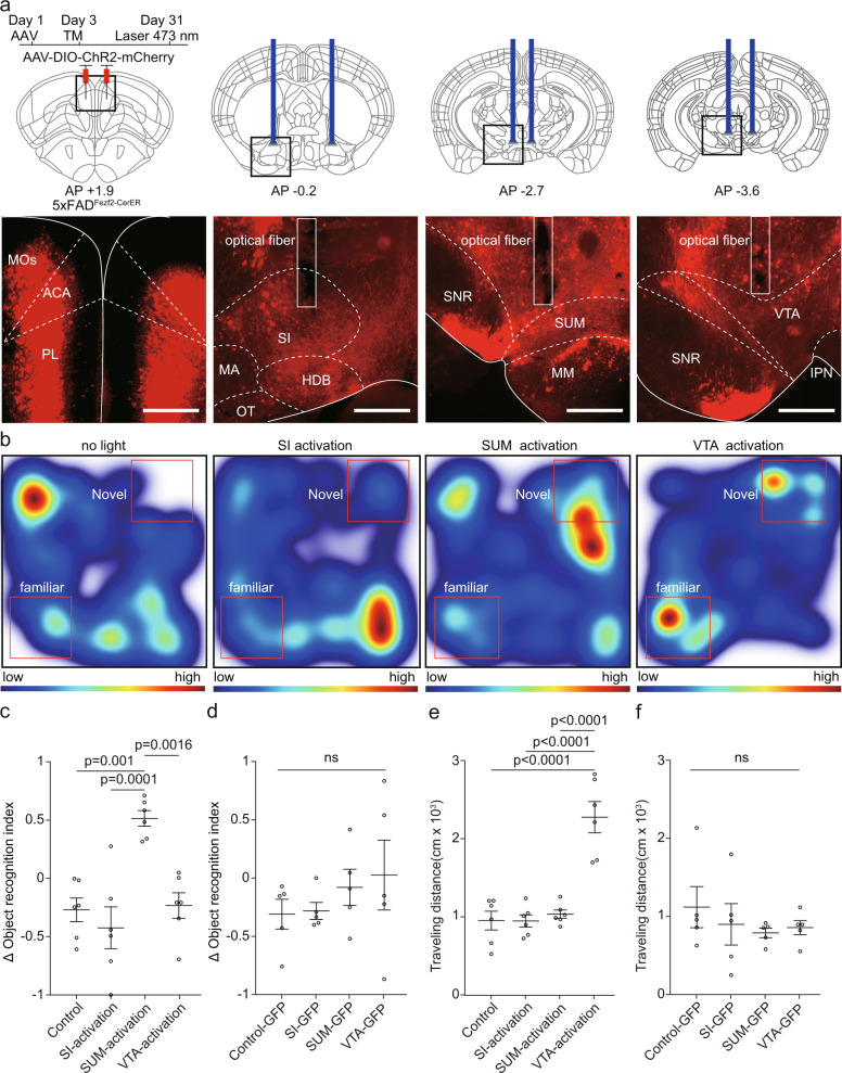
Fig. 3. Activation of specific axon terminals of extratelencephalic projection neurons in the mPFC of 5×FAD mice improved the object recognition memory expression.
a The experimental strategy of terminal activation of extratelencephalic projection neurons in the mPFC of 5×FAD mice. The optical fibers were implanted in SI, SUM, or VTA bilaterally to activate the axon terminals in these brain areas (top). The virus injection site and the positions of the optical fiber were shown below. b Heat-map plots showed object recognition memory measures from the object recognition test under different experimental conditions (no light stimulation; activation of axon terminals in SI; activation of axon terminals in SUM; activation of axon terminals in VTA.) Red = more time, blue = less time. c Statistical plots showed the delta object recognition index under different conditions in experimental group (mice that expressed ChR2 in the mPFC). one-way repeated-measures (RM) ANOVA with Tukey’s post hoc test, n = 6 animals. d Statistical plots showed the delta object recognition index under different conditions in control group (mice that expressed GFP in the mPFC). one-way repeated-measures (RM) ANOVA with Tukey’s post hoc test, n = 5 animals. e Statistical plots showed the traveling distance during the test session under different conditions in experimental group (mice that expressed ChR2 in the mPFC), one-way repeated-measures (RM) ANOVA with Tukey’s post hoc test, control vs SI -activation, p = 0.999995; control vs SUM-activation, p = 0.966536; control vs. VTA-activation, p = 0.000002; SI-activation vs. SUM-activation, p = 0.96159; SI-activation vs VTA-activation, p = 0.000002; SUM-activation vs. VTA-activation, p = 0.000005. n = 6 animals. f Statistical plots showed the traveling distance during the test session under different conditions in control group (mice that expressed GFP in the mPFC), one-way repeated-measures (RM) ANOVA with Tukey’s post hoc test, n = 5 animals. Scale bars in a are 500 μm. MOs secondary motor area, ACA anterior cingulate area, PL prelimbic area, SI substantia innominata, MA magnocellular nucleus, HDB horizontal diagonal band, OT olfactory tubercle, SNR substantia nigra, reticular part; SUM supramammillary nucleus, MM medial mammillary nucleus, VTA ventral tegmental area, IPN interpeduncular nucleus, TM tamoxifen. All data are listed as the Mean ± SEM.

