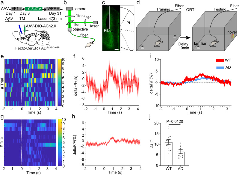
Fig. 6. Functional acetylcholine deficiency to extratelencephalic projection neurons in the mPFC of 5×FAD mice.
a Expressing the acetylcholine indicators in Fezf2 positive neurons in the mPFC with a Cre dependent virus. The Cre expression was induced by tamoxifen 3 days after the virus injection. b The scheme of fiber photometry. c The expression of acetylcholine indicators in Fezf2 positive neurons in the mPFC. d The experimental design of object recognition test. e Heatmap of acetylcholine response of extratelencephalic projection neurons in the PL of Fezf2-CreER mice during object interactions. f Average plots across different trials of individual animals of acetylcholine response of extratelencephalic projection neurons in PL of Fezf2-CreER mice during object interactions. g Heatmap of acetylcholine response of extratelencephalic projection neurons in PL of ADFezf2-CreER mice during object interactions. h Average plots across different trials of individual animals of acetylcholine response of extratelencephalic projection neurons in PL of ADFezf2-CreER mice during object interactions. i Average plots of acetylcholine response of extratelencephalic projection neurons in the PL of both Fezf2-CreER mice and ADFezf2-CreER mice during multiple tests on multiple animals (Fezf2-CreER, n = 12 animals; ADFezf2-CreER mice, n = 9 animals). j The quantification of AUC in i The acetylcholine response of extratelencephalic projection neurons in PL of both Fezf2-CreER mice is stronger than that in ADFezf2-CreER mice. (Two-tailed Mann-Whitney test, Fezf2-CreER mice, n = 12 animals; ADFezf2-CreER mice, n = 9 animals.) Scale bar in c is 200 μm. ACA anterior cingulate area, PL prelimbic area, TM tamoxifen. All data are listed as the Mean ± SEM. The color bars in the figure indicated df/f (%).

