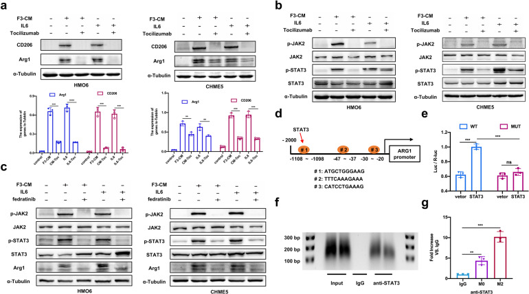
Fig. 5.
IL6/JAK2/STAT3 signaling mediates M2 polarization of microglia. a Western blot analysis of M2-marks (CD206 and Arg1) in HMO6 and CHME5 cells with control media or F3-CM alone or F3-CM treated with Tocilizumab (2.5 μg/ml for 24 h) or with IL6 (200 ng/ml for 24 h) alone or IL6-supplemented with Tocilizumab. b Western blot analysis of p-JAK2, JAK2, p-STAT3, and STAT3 in HMO6 and CHME5 cells with control media or F3-CM alone or F3-CM treated with Tocilizumab (2.5 μg/ml) or with IL6 (200 ng/ml) alone or IL6-supplemented with Tocilizumab. c Western blot analysis of CD206, Arg1, p-JAK2, JAK2, p-STAT3, and STAT3 in HMO6 and CHME5 cells with control media or F3-CM alone or F3-CM treated with fedratinib (20 μm for 24 h) or plus IL6(200 ng/ml) alone or IL6-supplemented with fedratinib. d The speculative binding sites in Arg1 for STAT3 were showed. e The dual-luciferase reporter experiment was applied to detect the luciferase activity of Arg1 stimulated by STAT3. f ChIP analysis was performed using a negative control immunoglobulin G (IgG) or anti-STAT3 antibody in CHME5 cells. g qPT-PCR of ChIP analysis was conducted to show binding site of STAT3 in ARG1. F3-CM: conditioned media of A549-F3 cells. Vector: pGL3-vector; STAT3: STAT3-vector. WT: wild type (#1); MUT: mutant type. M0:CHME5 cells; M2: F3-CM induced M2-CHME5 cells. Data are mean ± SD. *P < 0.05; **P < 0.01; ***P < 0.001

