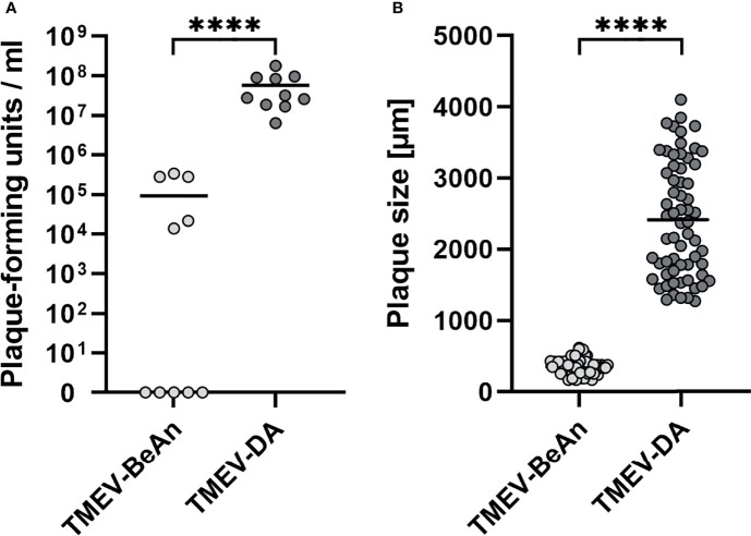
Figure 2.
Low numbers of infectious viral particles were found in IFN-β-/- mice after TMEV-BeAn infection, whereas TMEV-DA-infected IFN-β-/- mice developed a high viral load and had to be euthanized due to severe clinical disease (A). TMEV-BeAn and TMEV-DA induce small uniform plaques and large heterogeneous plaques, respectively (B). IFN-β-/- mice were intracranially infected with 1 x 10⁵ PFU of TMEV-BeAn or TMEV-DA. Plaque analysis was performed on L cells and used to detect infectious viral particles in the brain of TMEV-infected mice (A). Moreover, plaque sizes of TMEV strains used for mouse infection were determined (B). Mann-Whitney tests demonstrated statistically significant differences between the two TMEV strains (****p < 0.0001). Shown are all data points with means.

