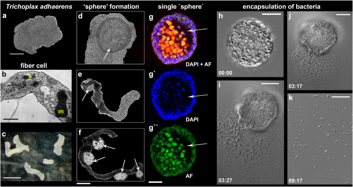
FIGURE 7.
“Sphere”-type formations in Trichoplax adhaerens. (A–C) control animals under optimal culture conditions (mode #1, see text and Figure 3); (B)—Transmission electron microscopy (TEM) image of the fiber cell with a bacterium (asterisk) and a large mitochondrial complex (m). (D–H)–Formation of spheres (arrows) from the upper/dorsal epithelium. (D)—disk-like animal; (E,F) -animals with elongated bodies; (E)—one “sphere” (arrow); (D)—four “spheres” (arrows). (G–G″)—Separated spheres with internal cavities (arrows); Nuclear DAPI staining—blue [excited by the violet (∼405 nm) laser with blue/cyan filter (∼460–470 nm)]; Autofluorescence (AF)—green (excitation 490 nm and emission 516 nm). (I–K)—Spherical formations encapsulate bacteria inside; (I–J)—the damage of the sphere’s surface by laser released numerous bacteria (magnified in (K); see text for details). Time intervals following the laser-induced injury are indicated in the left corners of each image. Scale: a 200 μm, (B)—500 nm, (C)—1 mm, (G–G″)—400 μm, (G–I)—20 μm, (K)—10 µm.

