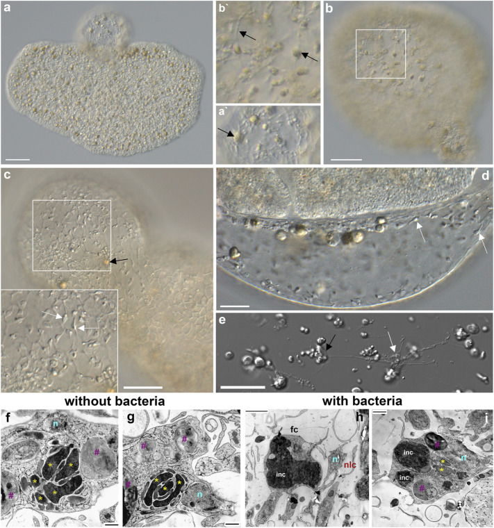
FIGURE 8.
Fiber cells in spherical structures (A–C) and their microanatomy (F–I). Both fiber cells (black arrows) and smaller neuroid-like cells (white arrows) are present in “spheres’”. (A)—Trichoplax adhaerens with a sphere on the upper side. A′– a part of the sphere with two fiber cells (arrow). (C,D,E)—smaller neuroid-like cells (white arrows) with elongated processes (E) that can form connections among themselves and fiber (black arrow) cells (Romanova et al., 2021). Both cell types are located in the middle layer of placozoans (B/B′). (F–I)—Transmission electron microscopy (TEM) of the fiber cells in Rickettsia-free Trichoplax (ampicillin-treated for 12 months) and control animals with endosymbiotic bacteria (see also Figure 7B). Of note, fiber cells in ampicillin-treated populations of placozoans had more elaborate mitochondrial clusters and clear inclusions. In contrast, in animals with bacteria, fiber cells possessed large dark (by TEM) inclusions (light microscopy also shows brownish inclusions—black arrows in A–C). Yellow asterisks (*) - mitochondria, n—nuclei, purple #—clear inclusions, inc—dark inclusions, fc—fiber cells, nlc—neuroid-like cells.

