Figure 2.
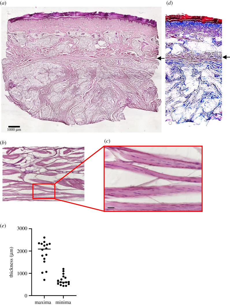
A layer of panniculus is found in heel tissues of human cadavers. (a) A cross-section of human cadaveric heel tissue stained with H&E. Panniculus carnosus is indicated by black arrow. Scale bar is 1000 µm. (b) A cross-section of human cadaveric heel tissue stained with Masson's trichrome reagent. Note the muscle fibres (black arrow) are stained red, while collagen is stained blue. (c,d) Close-ups of muscle fibres in the cadaveric heels. Scale bars are 50 µm. (e) Distribution of minimal and maximal thickness of panniculus layer in 16 cadaveric heel tissues (two samples from eight patients).

