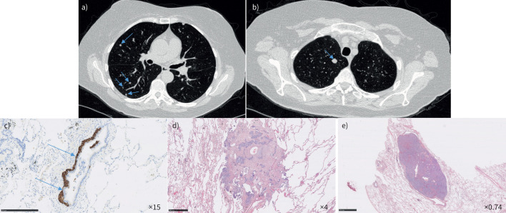
FIGURE 1.
Diffuse idiopathic pulmonary neuroendocrine cell hyperplasia in a 68-year-old woman. Chest computed tomography scan showed a) multiple nodules (arrows) and b) an 11-mm nodule in the right upper lobe (arrow). Wedge resection of the right upper lobe showed c) neuroendocrine cell hyperplasia beneath the bronchiolar epithelium (arrows) highlighted by chromogranin-A staining (scale bar=250 μm); d) a tumourlet (scale bar=500 μm); and e) a carcinoid tumour (haematoxylin and eosin staining) (scale bar=2.5 mm).

