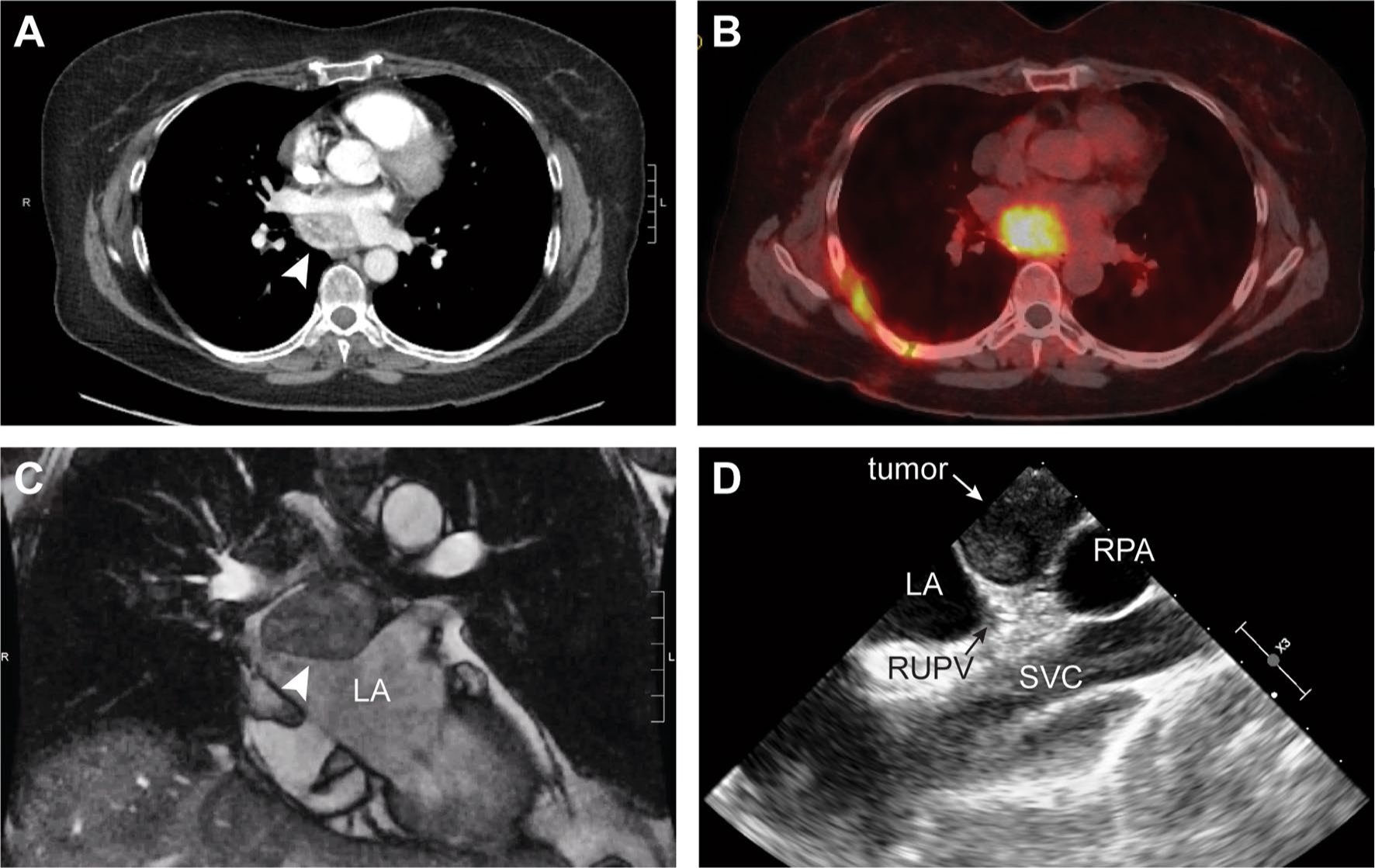
Fig. 1.

Pre-operative imaging diagnostics: a Computed tomography (CT) with IV contrast demonstrating a 4.7 × 3.4 cm enhancing mediastinal mass (arrow). b Positive Ga68-DOTATATE scan (SUV 13) demonstrating a single avid lesion with no distant disease, c Cardiac magnetic resonance imaging demonstrating mass (arrow) arising from the LA with arterial hyperenhancement and T2 hyperintensity, and d Transesophageal echo (TEE) demonstrating tumor with LA compression. LA, left atrium; RPA, right pulmonary artery; RUPV, right upper pulmonary vein; SVC, superior vena cava
