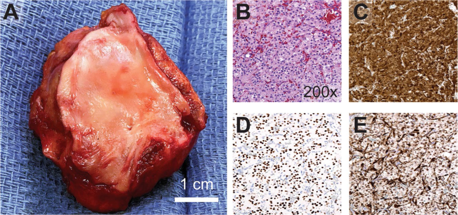
Fig. 3.

Pathologic assessment and immunohistochemistry: a Gross imaging of the specimen with en bloc portion of LA and underlying tumor mass. Histologic features including: b packed “Zellballen” growth of epithelioid tumor cells on hematoxylin and eosin stain, c strong and diffuse expression of chromogranin A, d INSM-1 stain highlighting chief cell nuclei, e S100 stain highlighting sustentacular cell nuclei at the periphery of cell nests (darkly staining)
