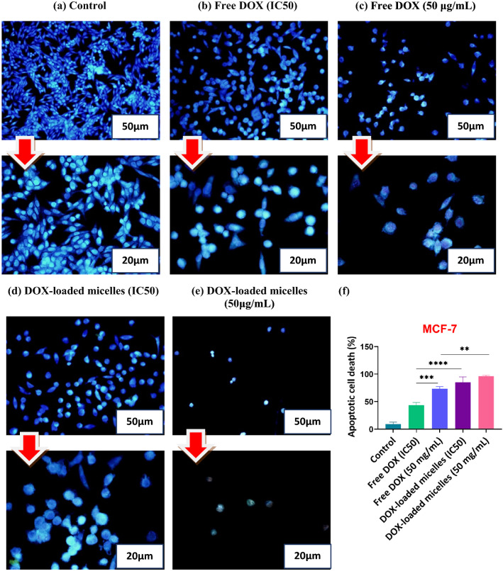
Fig. 7.
Fluorescence microscopic images of DAPI stained MCF-7 cells following 48 h of exposure to untreated cells (control) (a), free DOX (IC50) (b), free DOX (50 μg/mL) (c), DOX-loaded micelles (IC50) (d), and DOX-loaded micelles (50 μg/mL) (e). The percentages of apoptotic cell death in MCF-7 cells after being exposed to free DOX, and DOX-loaded micelles (f). As displayed in the diagram, DOX-loaded polymeric micelles yield highly considerable apoptosis (p < 0.01) in comparison to free DOX. Each value represents the mean value ± standard deviation (n = 3, *p < 0.05, ** p < 0.01, *** p < 0.001, ****p < 0.0001). Images were taken at magnification (×200) and (×400)

