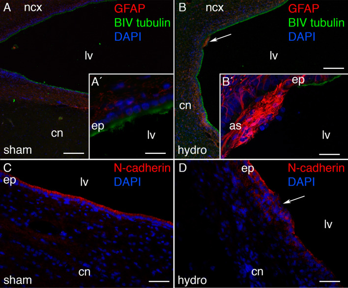
Fig. 2.
Ependymal disruption in the hydrocephalic pigs. Ependymal disruptions were detected in the hydrocephalic pigs through labeling the astrocytes (GFAP, fluorescence in red) and ependymal cells (βIV tubulin, green) in the periventricular area from A, A′ a sham control, and B a hydrocephalic pig. B′ Magnification of the top area pointed in B showing the ventricular zone disruption. C, D N-cadherin labelling also confirmed the disruption in the ventricular zone in the hydrocephalic pigs. Nuclear staining with DAPI in blue. Scale bars represent 120 µm in A, B; 25 µm in A′, and 40 µm in B′. Images were obtained under the confocal microscope, and a Z-stack of 10 µm was composed with ImageJ software. as astrocyte reaction, cn caudate nucleus, ep ependymal, ncx neocortex

