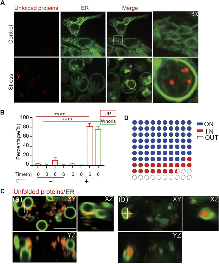
FIGURE 4.
ER whorls tether unfolded proteins under stress. (A) ER whorls and unfolded protein (UP) puncta appear only under stress. Scale bar = 10 μm. (B) Quantification of unfolded protein puncta and whorls under stress. A 6-h treatment of 10 mM DTT induced both unfolded protein and whorls formation at a final ratio of 81.7 ± 2.1% and 75.7 ± 2.0% (mean ± SD; from n = 235 cells), respectively. ****: p < 0.0001. (C) 3D view of colocalization of unfolded proteins and whorls. Unfolded proteins may be in contact with the outside surfaces of whorls (a) or exist inside the whorls (b). Scale bar = 1 μm. (D) Quantification of localization of unfold proteins with respect to whorls, 68.3 ± 0.3% (mean ± SD; from n = 355) of whorls have UPs attached to the membrane (ON), 19.3 ± 0.2% of whorls wrapped UPs inside (IN).

