
Fig. 4.
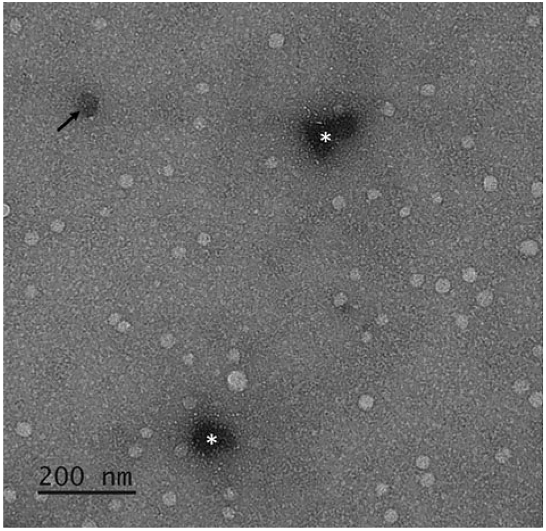
TEM image of exosomes. Due to the compartmentalized nature of exosomes, negative staining by UranyLess™ highlights intact bilipid layered vesicles as bright spherical spots relative to the darker, even background stained by UranyLess™. If an exosome is structurally compromised, it can appear as a darkened round spot as the stain is absorbed and retained within the vesicles (black arrow). Darker spots of undefined shapes are concentrations of stain (white asterisks). This phenomenon can be minimized by reducing the duration of time the sample is exposed to UranyLess™ but requires a balance as it would give a lighter background and may impair the contrast provided by the vesicles, which if present, are unstained